. 2010 Feb;151(2):502-12.
doi: 10.1210/en.2009-0678. Epub 2010 Jan 7.

Acute insulin signaling in pancreatic beta-cells is mediated by multiple Raf-1 dependent pathways

Affiliations

Acute insulin signaling in pancreatic beta-cells is mediated by multiple Raf-1 dependent pathways

Emilyn U Alejandro et al. Endocrinology. 2010 Feb.

Abstract

Insulin enhances the proliferation and survival of pancreatic beta-cells, but its mechanisms remain unclear. We hypothesized that Raf-1, a kinase upstream of both ERK and Bad, might be a critical target of insulin in beta-cells. To test this hypothesis, we treated human and mouse islets as well as MIN6 beta-cells with multiple insulin concentrations and examined putative downstream targets using immunoblotting, immunoprecipitation, quantitative fluorescent imaging, and cell death assays. Low doses of insulin rapidly activated Raf-1 by dephosphorylating serine 259 and phosphorylating serine 338 in human islets, mouse islets, and MIN6 cells. The phosphorylation of ERK by insulin was eliminated by exposure to a Raf inhibitor (GW5074) or transfection with a dominant-negative Raf-1 mutant. Insulin also enhanced the interaction between mitochondrial Raf-1 and Bcl-2 agonist of cell death (Bad), promoting Bad inactivation via its phosphorylation on serine 112. Insulin-stimulated ERK phosphorylation was abrogated by calcium chelation, calcineurin and calmodulin-dependent protein kinase II inhibitors, and Ned-19, a nicotinic acid adenine dinucleotide phosphate receptor (NAADPR) antagonist. Blocking Raf-1 and Ca(2+) signaling resulted in nonadditive beta-cell death. Autocrine insulin signaling partly accounted for the effects of glucose on ERK phosphorylation. Our results demonstrate that Raf-1 is a critical target of insulin in primary beta-cells. Activation of Raf-1 leads to both an ERK-dependent pathway that involves nicotinic acid adenine dinucleotide phosphate-sensitive Ca(2+) stores and Ca(2+)-dependent phosphorylation events, and an ERK-independent pathway that involves Bad inactivation at the mitochondria. Together our findings identify a novel insulin signaling pathway in beta-cells and shed light on insulin's antiapoptotic and mitogenic mechanisms.


Figures

Figure 1
Figure 1
Insulin promotes Raf-1 and ERK activation in mouse islets. A, Acute insulin signaling stimulation for 15 min in primary mouse islets with 0.2 and 200 nm insulin resulted in a loss of inhibitory phosphorylation of Raf-1 (pRaf-1) at serine 259. B, Mouse islets treated with 0.2 and 200 nm insulin for 30 min also caused an increase of the stimulatory phosphorylation of Raf-1 at serine 338 and a prosurvival phosphorylation of ERK (pErk; C). Bar graphs are quantification of Western blots using densitometry (n = 4 for mouse islets). *, Significant difference between treatment and serum-free control.
Figure 2
Figure 2
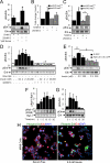
Insulin activates Raf-1 and ERK in transformed MIN6 cells. Panel A, ERK phosphorylation in MIN6 cells treated with low glucose (5 mm) or high glucose (25 mm) for 15 min with or without somatostatin (Soma, 1 μm; n = 3). Panel B, Insulin levels were measured in conditioned media of islets from Ins1−/−-Ins2+/+ mice treated with low and high glucose (n = 4). Panel C, ERK phosphorylation (pErk) in islets from Ins1−/−-Ins2+/− or Ins1−/−-Ins2+/+ mice or in response to 15 min treatment with 25 mm glucose or 5 mm glucose. Panels D and E, Insulin-induced ERK phosphorylation was blocked by a Raf inhibitor (E-GFP:GW5074; 10 μm; n = 4) or a dominant-negative Raf-1 (51-131)-GFP protein (n = 3). Panels F and G, Insulin (0.2 nm) caused an increase in Raf-1 and ERK phosphorylation in a time-dependent manner (n = 3). Panel H, Insulin induced phosphorylated ERK translocation in the nucleus of MIN6 cells (57,556 of 2,300,189 cells had nuclear ERK phosphorylation in control; 190,013 of 1,195,820 MIN6 cells had nuclear ERK phosphorylation with insulin). Bar graphs are quantification of Western blots using densitometry. *, Significant difference between treatment and control (serum free); ^, significant difference between insulin and insulin with GW5074 or dominant-negative Raf-1 (51-131)-GFP.
Figure 3
Figure 3
Insulin promotes Raf-1 translocation to the mitochondria. A, Insulin (0.2 nm) promoted Raf-1 mitochondrial localization compared with serum free in mouse pancreatic β-cell (n = 3). Scale bars, 10 μm. B, A plot of Pearson correlation r values between Raf-1 and Red-mitochondria tracker in mouse pancreatic β-cells treated in serum-free (SF) or 0.2 nm insulin condition. C, Phosphorylated Raf-1 (pRaf-1; serine 338), Raf-1, and cytochrome c oxidase IV (CoxIV) levels in total cell lysates or mitochondrial fraction of MIN6 cells treated with insulin for 10 min. Bar graph is a densitometry quantification (n = 3). *, Significant difference between treatment and control (serum free).
Figure 4
Figure 4
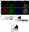
Endogenous Bcl-2 family members and Raf-1 form protein-protein interactions in pancreatic β-cell. A, Immunofluorescence imaging of endogenous Raf-1 and Bad in primary human and mouse β-cells. Pearson correlation r values between Raf-1 and Bad in human and mouse β-cells were 0.7 and 0.86, respectively. Scale bars, 10 μm. B, Immunoprecipitation (IP) demonstrating Raf-1 ability to form protein-protein interaction with Bcl-2 family proteins (Bcl-2, Bcl-xL, and Bad). IB, Immunoblot. C, Insulin increased Raf-1 and Bad protein-protein interaction (n = 3). D, Mouse islets treated with 0.2 and 200 nm insulin for 30 min caused an increase in the inhibitory phosphorylation of Bad (pBad) at serine 112 (n = 3). Bar graphs are quantification of Western blots using densitometry. *, Significant difference between treatment and serum-free control.
Figure 5
Figure 5
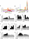
Ca2+ signaling modulators abrogates insulin-induced ERK activation. A, Ca2+ signals generated by insulin in individual dispersed human islet cells loaded with fura-4F in response to a range of insulin doses. B, Ca2+ traces of MIN6 cells loaded with fura-2 exposed to a ramp of insulin doses (550 cells imaged; 59 cells responded to insulin). Baseline solutions contained 3 mm glucose. C, MIN6 cells treated with insulin showed an increase on ERK phosphorylation (pErk) in 10 min that was blocked in the presence of BAPTA-AM. FBS, Fetal bovine serum. D, Phosphorylated ERK levels in MIN6 cells treated with GW5074 (10 μm) and BAPTA-AM (50 μm) individually or both for 6 h (n = 3). E, Phosphorylated Bad (serine 112) levels in MIN6 cells treated with GW5074 and BAPTA-AM individually or both for 6 h (n = 3). Rapid increase in PI incorporation in dispersed mouse islet cells (F) and MIN6 cells (G) treated with GW5074, BAPTA-AM, or both (n = 6). Area under the curve (AUC) of PI incorporation from 1 to 48 h after treatment. H, Cleaved caspase-3 expression levels in MIN6 cells treated with GW5074 and BAPTA-AM for 6 h (n = 3). I–K, MIN6 cells treated with insulin show an increase in ERK phosphorylation that was blocked in the presence of Ned-19 (I, NAADP receptor antagonist), KN-93 (J, CaMKII inhibitor), and FK506 (K, calcineurin inhibitor) for 10 min treatment (n = 3). Bar graphs are quantification of Western blots using densitometry. *, Significant difference (P < 0.05) between the serum-free control and treatment; ^, significant difference between insulin and insulin with inhibitors: BAPTA-AM (50 μm), Ned-19 (100 μm), KN-93 (1 μm), and FK506 (50 nm).

References

    1. Johnson JD, Bernal-Mizrachi E, Alejandro EU, Han Z, Kalynyak TB, Li H, Beith JL, Gross J, Warnock GL, Townsend RR, Permutt MA, Polonsky KS 2006 Insulin protects islets from apoptosis via Pdx1 and specific changes in the human islet proteome. Proc Natl Acad Sci USA 103:19575–19580 - PMC - PubMed
    1. Ueki K, Okada T, Hu J, Liew CW, Assmann A, Dahlgren GM, Peters JL, Shackman JG, Zhang M, Artner I, Satin LS, Stein R, Holzenberger M, Kennedy RT, Kahn CR, Kulkarni RN 2006 Total insulin and IGF-I resistance in pancreatic β cells causes overt diabetes. Nat Genet 38:583–588 - PubMed
    1. Okada T, Liew CW, Hu J, Hinault C, Michael MD, Krtzfeldt J, Yin C, Holzenberger M, Stoffel M, Kulkarni RN 2007 Insulin receptors in β-cells are critical for islet compensatory growth response to insulin resistance. Proc Natl Acad Sci USA 104:8977–8982 - PMC - PubMed
    1. Bernal-Mizrachi E, Fatrai S, Johnson JD, Ohsugi M, Otani K, Han Z, Polonsky KS, Permutt MA 2004 Defective insulin secretion and increased susceptibility to experimental diabetes are induced by reduced Akt activity in pancreatic islet β cells. J Clin Invest 114:928–936 - PMC - PubMed
    1. Hashimoto N, Kido Y, Uchida T, Asahara S, Shigeyama Y, Matsuda T, Takeda A, Tsuchihashi D, Nishizawa A, Ogawa W, Fujimoto Y, Okamura H, Arden KC, Herrera PL, Noda T, Kasuga M 2006 Ablation of PDK1 in pancreatic β cells induces diabetes as a result of loss of β cell mass. Nat Genet 38:589–593 - PubMed

Publication types

MeSH terms