

Involvement of CYR61 and CTGF in the fascin-mediated proliferation and invasiveness of esophageal squamous cell carcinomas cells
- PMID: 20056838
- PMCID: PMC2808098
- DOI: 10.2353/ajpath.2010.090118
Involvement of CYR61 and CTGF in the fascin-mediated proliferation and invasiveness of esophageal squamous cell carcinomas cells
Erratum in
- Am J Pathol. 2010 May;176(5):2581
Abstract
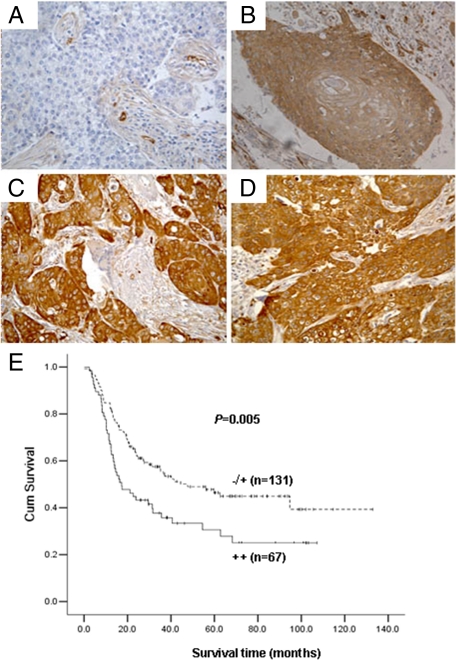
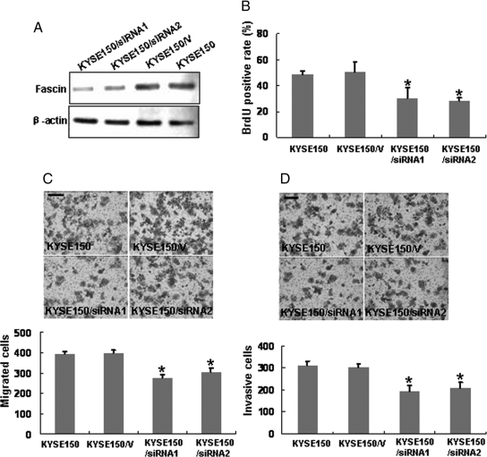
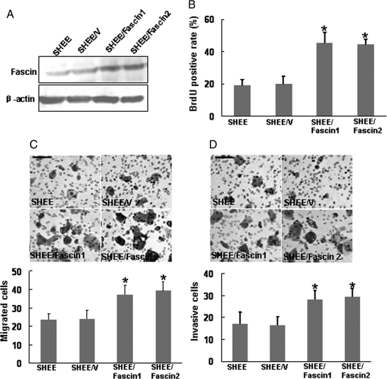
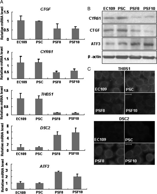
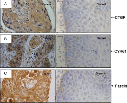
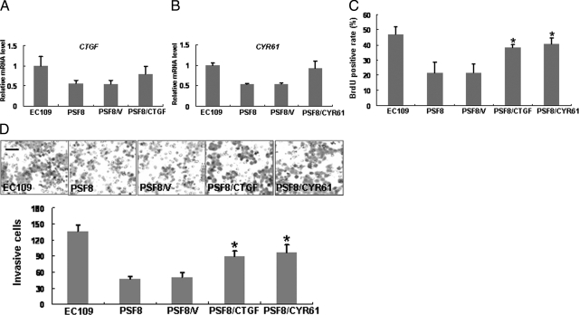
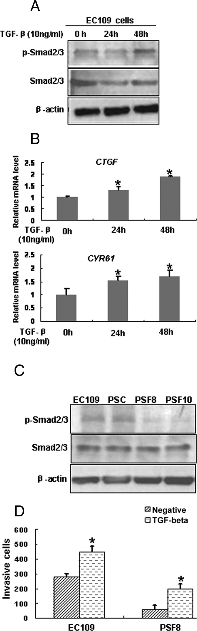
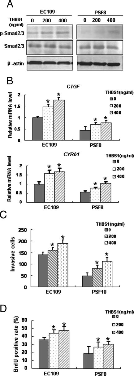
Fascin is overexpressed in esophageal squamous cell [corrected] carcinoma (ESCC) and involved in the proliferation and invasiveness of ESCC cells. In this study, we retrospectively examined the expression of fascin in ESCC samples by immunohistochemistry and revealed that overexpression of fascin was related to poor patient survival. RNAi-mediated knockdown of fascin in ESCC cells significantly inhibited cell proliferation and invasiveness, whereas forced expression of fascin in immortalized esophageal epithelial cells accelerated cell proliferation and invasiveness. To explore the underlying mechanism, cDNA microarray was performed to identify the differential gene expression profiles between a fascin-depleted cell line by RNAi and the corresponding control ESCC cells. Results showed that 296 genes were differentially expressed on fascin depletion. In this study, we focused on two down-regulated genes: CYR61 and CTGF. We found that restored expression of either CYR61 or CTGF led to a recovery of the suppression of cellular proliferation and invasiveness induced by down-regulation of fascin expression; the protein level of CYR61 and CTGF were up-regulated in ESCCs and their expression pattern correlated with fascin overexpression. Finally, analysis of signal transduction revealed that fascin affected the expressions of CYR61 and CTGF through transforming growth factor (TGF)-beta pathway. Taken together, we propose that fascin regulates the proliferation and invasiveness of ESCC cells by modulating the expression of CTGF and CYR61 via TGF-beta pathway.
Figures
Similar articles
-
Expression and prognostic significance of THBS1, Cyr61 and CTGF in esophageal squamous cell carcinoma.BMC Cancer. 2009 Aug 22;9:291. doi: 10.1186/1471-2407-9-291. BMC Cancer. 2009. PMID: 19698122 Free PMC article.
-
Roles of ezrin in the growth and invasiveness of esophageal squamous carcinoma cells.Int J Cancer. 2009 Jun 1;124(11):2549-58. doi: 10.1002/ijc.24216. Int J Cancer. 2009. PMID: 19165868
-
Prognostic significance of fascin overexpression in human esophageal squamous cell carcinoma.Clin Cancer Res. 2005 Apr 1;11(7):2597-605. doi: 10.1158/1078-0432.CCR-04-1378. Clin Cancer Res. 2005. PMID: 15814639
-
Role of fascin in the proliferation and invasiveness of esophageal carcinoma cells.Biochem Biophys Res Commun. 2005 Nov 11;337(1):355-62. doi: 10.1016/j.bbrc.2005.09.055. Biochem Biophys Res Commun. 2005. PMID: 16185662
-
Cyr61/CTGF/Nov family proteins in gastric carcinogenesis.World J Gastroenterol. 2014 Feb 21;20(7):1694-700. doi: 10.3748/wjg.v20.i7.1694. World J Gastroenterol. 2014. PMID: 24587648 Free PMC article. Review.
Cited by
-
F806 Suppresses the Invasion and Metastasis of Esophageal Squamous Cell Carcinoma via Downregulating F-Actin Assembly-Related Rho Family Proteins.Biomed Res Int. 2018 Sep 19;2018:2049313. doi: 10.1155/2018/2049313. eCollection 2018. Biomed Res Int. 2018. PMID: 30327774 Free PMC article.
-
A novel staging model to classify oesophageal squamous cell carcinoma patients in China.Br J Cancer. 2014 Apr 15;110(8):2109-15. doi: 10.1038/bjc.2014.101. Epub 2014 Feb 25. Br J Cancer. 2014. PMID: 24569468 Free PMC article.
-
Macrolide analog F806 suppresses esophageal squamous cell carcinoma (ESCC) by blocking β1 integrin activation.Oncotarget. 2015 Jun 30;6(18):15940-52. doi: 10.18632/oncotarget.3612. Oncotarget. 2015. PMID: 25909284 Free PMC article.
-
Epigenetic silencing of HOPX promotes cancer progression in colorectal cancer.Neoplasia. 2012 Jul;14(7):559-71. doi: 10.1593/neo.12330. Neoplasia. 2012. PMID: 22904674 Free PMC article.
-
SREBP2 is upregulated in esophageal squamous cell carcinoma and co‑operates with c‑Myc to regulate HMGCR expression.Mol Med Rep. 2019 Oct;20(4):3003-3010. doi: 10.3892/mmr.2019.10577. Epub 2019 Aug 9. Mol Med Rep. 2019. PMID: 31432128 Free PMC article.
References
-
- Mariette C, Balon JM, Piessen G, Fabre S, Van Seuningen I, Triboulet JP. Pattern of recurrence following complete resection of esophageal carcinoma and factors predictive of recurrent disease. Cancer. 2003;97:1616–1623. - PubMed
-
- Law SY, Fok M, Cheng SW, Wong J. A comparison of outcome after resection for squamous cell carcinomas and adenocarcinomas of the esophagus and cardia. Surg Gynecol Obstet. 1992;175:107–112. - PubMed
-
- Ding Y, Shimada Y, Gorrin-Rivas MJ, Itami A, Li Z, Hong T, Maeda M, Komoto I, Kawabe A, Kaganoi J, Imamura M. Clinicopathological significance of human macrophage metalloelastase expression in esophageal squamous cell carcinoma. Oncology. 2002;63:378–384. - PubMed
-
- Adams JC. Formation of stable microspikes containing actin and the 55 kDa actin bundling protein, fascin, is a consequence of cell adhesion to thrombospondin-1: implications for the anti-adhesive activities of thrombospondin-1. J Cell Sci. 1995;108:1977–1990. - PubMed
Publication types
MeSH terms
Substances
LinkOut - more resources
Full Text Sources
Medical
Molecular Biology Databases
Miscellaneous
