Comparative Study
. 2010 Feb;176(2):660-78.
doi: 10.2353/ajpath.2010.090348. Epub 2010 Jan 7.

Weaving hypothesis of cardiomyocyte sarcomeres: discovery of periodic broadening and narrowing of intercalated disk during volume-load change

Affiliations
Comparative Study

Weaving hypothesis of cardiomyocyte sarcomeres: discovery of periodic broadening and narrowing of intercalated disk during volume-load change

Makoto Yoshida et al. Am J Pathol. 2010 Feb.

Abstract

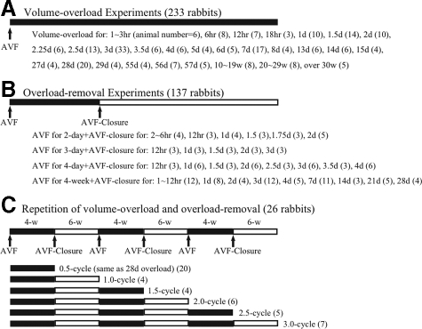
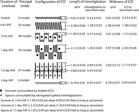
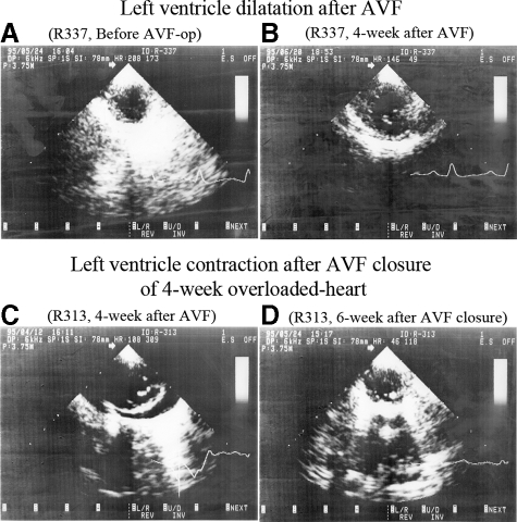
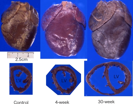
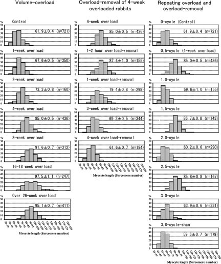
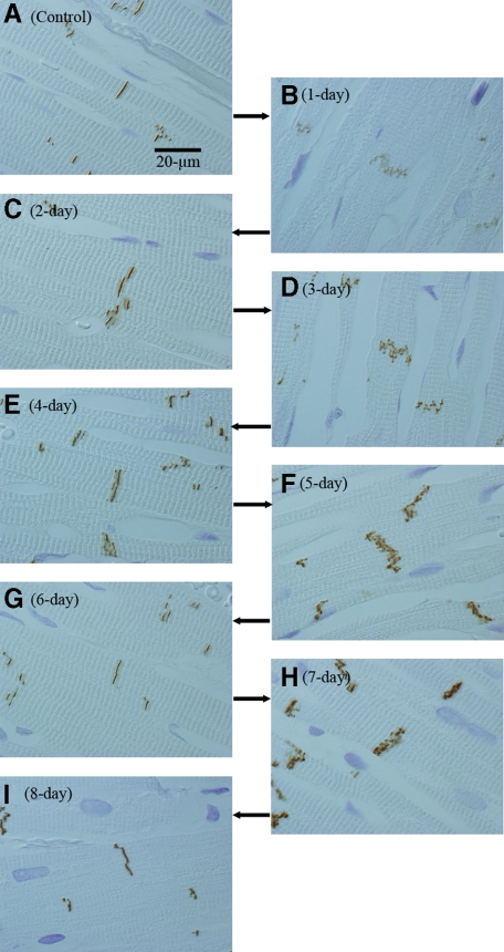
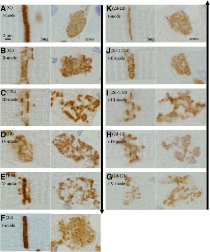
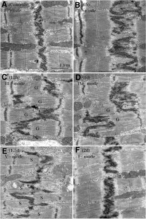
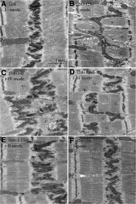
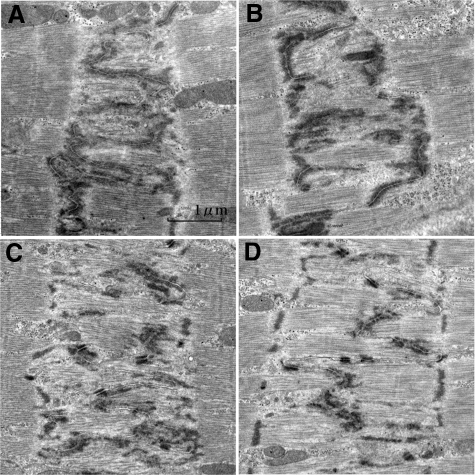
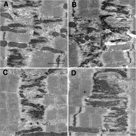
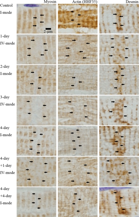
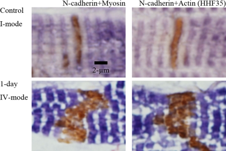
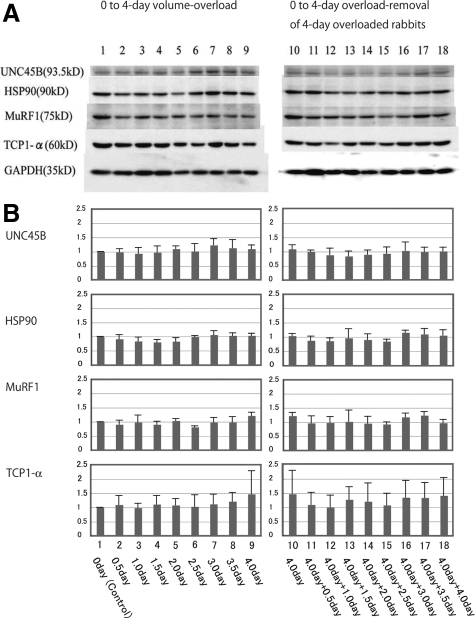
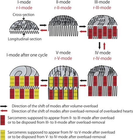
To investigate how cardiomyocytes change their length, echocardiographic and morphological studies were performed on rabbit hearts that were subjected to volume overload, overload removal, and repeated cycles of overload and overload removal. These conditions were created by arterio-venous fistula between the carotid artery and jugular vein, closure of the fistula, and cycles of repeatedly forming and closing fistula, respectively. After overload, hearts dilated and myocytes elongated. Intercalated disks repeatedly broadened and narrowed with a 2-day cycle, which continued for 8 weeks in many animals. The cycle consisted of shifts between five modes characterized by two interdigitation elongation-and-shortenings as follows: (I) flat with short ( approximately 1/4 to approximately 1/3 sarcomere long) interdigitations; (II) flat with long (one sarcomere long) interdigitations; (III) grooved with short interdigitations; (IV) grooved with long interdigitations; (V) flat with short interdigitations intermingled by sporadic long interdigitations; and return to (I). After overload removal, hearts contracted and myocytes shortened with similar 2-day broadening and narrowing cycle of intercalated disks, in which the five modes were reversed. Repeated overload and overload removal resulted in the repetition of myocyte elongation and shortening. We hypothesize that a single elongation-and-shortening event creates or disposes one sarcomere layer, and the two consecutive elongation-and-shortenings occur complementarily to each other so that the disks return to their original state after each cycle. Our hypothesis predicts that intercalated disks weave and unravel one sarcomere per myocyte per day.


Figures

Figure 1
Figure 1
Experimental schedule.
Figure 2
Figure 2
Relation between left ventricular long axis-to-short axis ratio and short axis lumen diameter.
Figure 3
Figure 3
Relation between left ventricular volume and short axis lumen diameter.
Figure 4
Figure 4
Schematic illustration of intercalated disk configuration and measurements of interdigitation length and intercalated disk width during 2 days after volume overload.
Figure 5
Figure 5
Echocardiographic images after volume overload and overload removal. Left ventricle dilates from A to B by 4-week AVF. Four-week overloaded heart contracts from C to D by 6-week AVF closure. Left ventricle cross-sectional planes at end-diastolic phase (B-mode echocardiography).
Figure 6
Figure 6
Gross appearance of volume-overloaded hearts. Hearts show dilatory hypertrophy after volume overload. Left: Control, Middle: 4-week after AVF, Right: 30-week after AVF.
Figure 7
Figure 7
Histogram of myocyte length. The left column is histograms of volume overload experiments. Histograms shift to the right gradually until 4 weeks. The myocyte length (ML) at 4 weeks is significant longer than that of 2 weeks (P < 0.0001), which is significantly longer than that of control (P < 0.0001). At 8 weeks, ML is significantly longer than that of 4 weeks and some long myocytes reach 140 sarcomeres. At 16 to 18 weeks and over 26 weeks, the ML is significantly longer than that of 8 weeks (P < 0.0001) and very long myocytes reach 180 sarcomeres. The middle column is histograms of overload-removal experiments of 4-week overloaded animals. Histograms shift to the left gradually until 4-week overload removal. The ML of 4-week overload removal is significantly shorter than that of 3-week overload removal (P < 0.0001), which is significantly shorter than that of 1-week overload removal (P < 0.0001). ML of 1-week overload removal is significantly shorter than that of 1- to 2-hour overload removal (P < 0.0001) or that of 4-week overloaded animals (P < 0.0001). The right column is histograms of repeating overload and overload-removal. Histograms of 1.0-cycle, 2.0-cycle and 3.0-cycle are similar to histogram of control, while those of 1.5-cycle and 2.5-cycle are similar to histogram of 4-week overload (0.5-cycle). ML of 1.5-cycle and 2.5-cycle are significantly longer than those of 1.0-cycle, 2.0-cycle, 3.0-cycle and 3-cycle sham, respectively (P < 0.0001). ML is expressed mean ± SEM in each histogram. (n) is number of measured myocytes. X bar indicates sarcomere number in 10-sarcomere steps and Y bar indicates % of myocytes.
Figure 8
Figure 8
Broadening and narrowing of intercalated disks after volume overload. In controls, ICDs are narrow and simple (A). After 1 (B), 3 (D), 5 (F) and 7-day (H) volume overload, ICDs are broad, while ICDs are narrow after 2 (C), 4 (E), 6 (G), and 8-day (I) volume overload. ICDs are stained by N-cadherin. Images are obtained with differential microscopy to define sarcomeres.
Figure 9
Figure 9
Microscopic appearances of the shift of five modes of intercalated disk after overload and after overload removal. Left column (volume overload): In the control, ICD is narrow and straight in longitudinal-section (long) and flat and simple in cross section (cross) (A[C]; Control; I-mode). After 6-hour (B [6 hours]), ICD is almost one-sarcomere-wide with elongated interdigitations in longitudinal section and flat and simple in cross section (II-mode). After 12-hour (C [12 hours]) ICD separates into two lines with a distance of one-sarcomere-stretch between them. Each line is narrow with short interdigitations. Cross section reveals irregular worm-eaten pattern indicating that disks are filled with one-sarcomere-deep grooves and contra-grooves (III-mode). After 1 day (D [1 day]), ICDs broaden. Disks are filled with one-sarcomere-deep grooves plus contra-grooves, the bottoms of which consist of elongated interdigitations resulting in ∼two-sarcomeres-wide ICD (IV-mode). After 1.5 days (E [1.5 days]), the ICD is flat and thin, but sporadic one-sarcomere-long interdigitations appear as spikes (V-mode). After 2 days (F [2 days]), ICDs are narrow and simple like those of control (I-mode). Right column (overload removal of 2-day overloaded hearts): After 12-hour (G [2 days−12 hours]), the ICD is flat and thin, but sporadic one-sarcomere-long interdigitations appear as spikes (r-V-mode). After 1 day (H [2 days − 1 day]), the ICD broadens. It is filled with one-sarcomere-deep grooves plus contra grooves, the bottoms of which consist of elongated interdigitations, resulting in two-sarcomeres-wide ICD (r-IV-mode). After 1.5 days (I [2 days −1.5 days]), the ICD separates into two lines with a distance of one-sarcomere stretch between them. Each line is narrow with short interdigitations. A cross section reveals irregular worm-eaten pattern showing that disks are filled with one-sarcomere-deep grooves and contra-grooves (r-III-mode). After 1.75 days (J [2 days −1.75 days]), the ICD is almost one-sarcomere-wide with elongated interdigitations so that cross section reveals a simple flat disk. These represent flat thick disks (r-II-mode). After 2 days (K [2 days − 2 days]), the ICD is narrow and simple like those of the control (r-I-mode). Modes appeared in overload removal are similar and indistinguishable from those that appeared after overload. The ICDs are stained with N-cadherin. The arrow in the left lane (upper to lower) indicates time course direction of overload and the arrow in the right lane (lower to upper) indicates time course of overload removal.
Figure 10
Figure 10
Ultrastructural changes of the intercalated disk after volume overload. The control shows thin and flat disks with short interdigitations (1/4- to 1/3-sarcomere long) (A [Control]) (I-mode). At 6 hours of volume-overload, the ICD becomes thick with one-sarcomere-long interdigitations (B [6 hours]) (II-mode). At 12 hours, the ICD is folded to form ∼one-sarcomere-deep grooves (G) and contra-grooves (G) with short interdigitations (C [12 hours]) (III-mode). At 1 day, interdigitations elongate up to one sarcomere long in the folded ICD (D [1 day]) (IV-mode) so that the ICD broadens to ∼two sarcomeres wide. Grooves (G) and contra-grooves (G) appear in this mode. At 1.5 days, the ICD is thin with mostly short interdigitations, but one-sarcomere-long interdigitations sporadically appear as spikes (E [1.5 days]) (V-mode). There appear “spaces” (S) surrounded by several spikes. At 2 days, the ICD is thin and flat with short interdigitations similar to those of controls, finishing one cycle of ICD changes during volume overload (F [2 days]) (I-mode). Longitudinal section to myocytes.
Figure 11
Figure 11
Ultrastructural changes of the intercalated disk of 2-day overloaded hearts followed by overload removal. A 2-day overloaded heart shows thin and flat disks as observed in Figure 10 (A [2 days]) (I-mode). At 12 hours of overload removal, the ICD is thin with mostly short interdigitations, but there appear sporadic one-sarcomere-long interdigitations (B [2 days + 12 hours]) (r-V-mode). There appear “spaces” (S) surrounded by several spikes. At 1 day of overload removal, interdigitations elongate up to one sarcomere long bilaterally so that the ICD is folded to form one-sarcomere-deep grooves (G) and contra-grooves (G). Elongated interdigitation plus grooves results in ∼two-sarcomere-wide broad ICDs (C [2 days + 1 day]) (r-IV-mode). At 1.5 days of overload removal, the ICD is folded with short interdigitations keeping one-sarcomere-deep grooves and contra-grooves (G) (D [2 days + 1.5 days]) (r-III-mode). At 1.75 days of overload removal, the ICD is flat with one-sarcomere-long interdigitations (E [2 days + 1.75 days]) (r-II-mode). At 2 days of overload-removal, the ICD is thin and flat with short interdigitations, finishing one cycle of ICD changes during overload removal (F [2 days + 2 days]) (r-I-mode). Modes appeared in overload removal are similar and indistinguishable from those after overload. Longitudinal section to myocytes.
Figure 12
Figure 12
Intercalated disks of assumed transitional II-mode to III-mode and IV-mode to V-mode after overload. Assumed transitional II to III-mode (A, B): In (A) (6-hour overload), the ICD is one sarcomere wide. Profiles of interdigitations are obscure in some portions. Filaments appear to form bundle in the areas of widened ICD, but they show no Z-band. In (B) (6-hour overload), there are filament bundles with plausible Z-band in the areas of widened ICD. Assumed transitional IV to V-mode (C, D): In (C) (1-day overload), ICD is nearly two sarcomeres wide. Interdigitations are complicated. Filaments within ICDs are going to form bundle formation, but they show no distinct Z-band. In (D) (1-day overload), ICD is two sarcomeres wide. Interdigitations are mostly in the middle line, but some elongate one-sarcomere-long bilaterally like V-mode. Filaments between interdigitation form bundles with Z-band. They would be immature stage of newly formed sarcomeres. Longitudinal section to myocytes.
Figure 13
Figure 13
Intercalated disks of assumed transitional r-V-mode to r-IV-mode and r-III-mode to r-II-mode after overload removal. Assumed transitional r-V-mode to r-IV-mode (A, B): In (A) (2-day overload and 12-hour overload removal), there appear foci with no filament in the spaces surrounded by elongated interdigitations. In (B) (2-day overload and 12-hour overload removal), some foci of no filament appeared, where irregularly elongated interdigitations were found. Assumed transitional r-III-mode to r-II-mode (C, D): In (C) (2-day overload and 1.5-day overload removal), there appear foci with no filament within the widened ICD. In (D) (2-day overload and 1.5-day overload removal), irregular and monotonously electron dense portions appeared within widened ICDs, where intercellular junctions become obscure. Appearance of no filament zones would be ongoing stage of sarcomere disposal. Longitudinal section to myocytes.
Figure 14
Figure 14
Myosin, actin, and desmin around intercalated disk in I-mode and IV-mode. Myosin, actin (HHF35), and desmin are individually immunohistochemically stained with paraffin section (nonperfused fixed hearts). They are regularly stained in the myocyte body; however, around ICDs, which we suppose to exist in the zones indicated by arrows, myosin, actin, and desmin show characteristic features as follows. In I-mode [control, 2-day after AVF (2-day), 4-day after AVF (4-day) and 4-day after overload removal of 4-day overloaded animal (4-day + 4-day)], the zones are narrow and simple. They are interposed by two myosin stained lines and two desmin stained lines. They are vaguely stained by actin. In IV-mode [1-day after AVF (1-day), 3-day after AVF (3-day), 1-day after overload-removal of 4-day overloaded animal (4-day + 1-day)], the zones are wide and irregular. Nearly two sarcomere-wide zones appear between myosin stained lines. Actin is irregular and vague and desmin is irregular in these zones.
Figure 15
Figure 15
Double immunohistochemical observation of intercalated disk at I and IV-mode. N-cadherin and myosin (N-cadherin+myosin) and N-cadherin and actin (N-cadherin+HHF35) are double immunohistochemically stained in control (I-mode) and 1 day after AVF (IV-mode). N-cadherin stained ICD is narrow and simple interposed between two myosin lines and actin lines in I-mode. N-cadherin stained ICD is wide and irregular in IV-mode. The zone margined by myosin or actin is filled with irregular and widened ICDs. Some actin positive portions appear within ICDs.
Figure 16
Figure 16
Immunofluorescent images of major filaments (actin, myosin, and titin), chaperons (TCP-1α, HSP90 and UNC45B) and ubiquitin ligase (MuRF1). Upper panel: Actin (phalloidin), myosin, and titin are observed (frozen section, 1-day after AVF of 4-day overloaded-heart; IV-mode). These filaments are regularly arranged in the body of myocytes, while ICDs could not be well identified so long as we observed. Lower panel: Chaperon and ubiquitin ligase (TCP-1α, HSP90, UNC45B, MuRF1) are observed (paraffin section, 1.5-day after AVF; V-mode). Their activity could not be identified. Frozen section also expresses no identifiable activity of these proteins. Laser confocal microscopy.
Figure 17
Figure 17
Western blot of chaperons (TCP-1α, HSP90 and UNC45B) and ubiquitin ligase (MuRF1) in volume-overloaded hearts and overload-removed hearts. Left panel shows overload experiments; Lane 1: Control (0-day), Lane 2: 0.5-day AVF, Lane 3: 1-day AVF, Lane 4: 1.5-day AVF, Lane 5: 2-day AVF, Lane 6: 2.5-day AVF, Lane 7: 3-day AVF, Lane 8: 3.5-day AVF, Lane 9: 4-day AVF. Right panel shows overload-removal experiments of 4-day overloaded rabbits; Lane 10: 4-day AVF, Lane 11: 4-day AVF plus 0.5-day AVF closure, Lane 12: 4-day AVF plus 1-day AVF closure, Lane 13: 4-day AVF plus 1.5-day AVF closure, Lane 14: 4-day AVF plus 2-day AVF closure, Lane 15: 4-day AVF plus 2.5-day AVF closure, Lane 16: 4-day AVF plus 3-day AVF closure, Lane 17: 4-day AVF plus 3.5-day AVF closure, Lane 18: 4-day AVF plus 4-day AVF closure. Upper panel (A) shows one representative Western blot of three chaperons (UNC45B, HSP90, and TCP1-α) and one ubiquitin ligase (MuRF1). Lower panel (B) shows graphs of ratio to control of these proteins. Error bar indicates mean ± SEM. Three chaperons and ubiquitin ligase shows no significant difference among overload experiments. They show no significant difference among overload removal experiments. They show no significant difference between the group of overload experiments and that of overload removal experiments.
Figure 18
Figure 18
Hypothetical shift of five modes of intercalated disks after overload and overload removal. Volume overload: During volume overload, modes shift from I to V and then returned to I, finishing one cycle of ICD changes (black arrows). From I-mode to II-mode interdigitations elongate one sarcomere. From II-mode to III-mode, two adjacent interdigitations alternately retract to form folds, which are grooves and contra-grooves three-dimensionally. Within folds, new one-sarcomere-long myofibril bundles (red) appear. Observing from the other side, top of the grooves consists of three adjacent interdigitations. From III-mode to IV-mode, interdigitations again elongate. From IV-mode to V-mode, one center interdigitation among three continual interdigitations retracts. Laterally located interdigitations remain elongated, which resemble spikes. Within spaces, surrounded laterally by spiked interdigitations and bottomed with retracted interdigitation, new one-sarcomere-long myofibril bundles (yellow) appear. Because the second retraction is complementary to the first, new myofibril bundles in the second retraction are alternately placed between those in the first retraction. From V-mode to I-mode, spiked-interdigitations retract. After one cycle, two layers of sarcomere are created at each side of the ICDs. Overload-removal: After overload-removal of 2-day (or “even” number day) overloaded hearts, modes shift from I-mode after cycle, r-V, r-IV, r-III, r-II to r-I-mode (I-mode after reverse cycle) (red arrows). First, interdigitations elongate one-sarcomere-long sporadically and bilaterally at first (I to r-V-mode). Then, short interdigitations between sporadically elongated interdigitations elongate bilaterally to dispose one-sarcomere-long myofibril bundles (yellow) (r-V to r-IV-mode). Elongated interdigitations shorten from r-IV to r-III-mode. Two interdigitations in the bottoms again elongate into the grooves to dispose one-sarcomere-long myofibril bundles (red) (r-III to r-II-mode). Because this elongation is exactly complementary to the first elongation, interdigitations line straight. Elongated interdigitations shorten from r-II to r-I-mode. This five mode shifts are exactly reverse manner to those after overload so that two layers of sarcomere are disposed at each side of the ICDs.

Similar articles

Cited by

References

    1. Linzbach AJ. Heart failure from the point of view of quantitative anatomy. Am J Cardiol. 1960;5:370–382. - PubMed
    1. Ross J., Jr Adaptations of the left ventricle to chronic volume overload. Circ Res. 1974;35 suppl II:64–70. - PubMed
    1. Hunter JJ, Chien KR. Signaling pathways for cardiac hypertrophy and failure. New Eng J Med. 1999;341:1276–1283. - PubMed
    1. Gerdes AM, Campbell SE, Hilbelink DR. Structural remodeling of cardiac myocytes in rats with arteriovenous fistulas. Lab Invest. 1988;59:857–861. - PubMed
    1. Huang M, Hester RL, Guyton AC. Hemodynamic changes in rats after opening an arteriovenous fistula. Am J Physiol. 1992;262:H846–H851. - PubMed

Publication types

MeSH terms