

A transient niche regulates the specification of Drosophila intestinal stem cells
- PMID: 20056890
- PMCID: PMC2857772
- DOI: 10.1126/science.1181958
A transient niche regulates the specification of Drosophila intestinal stem cells
Abstract
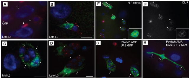
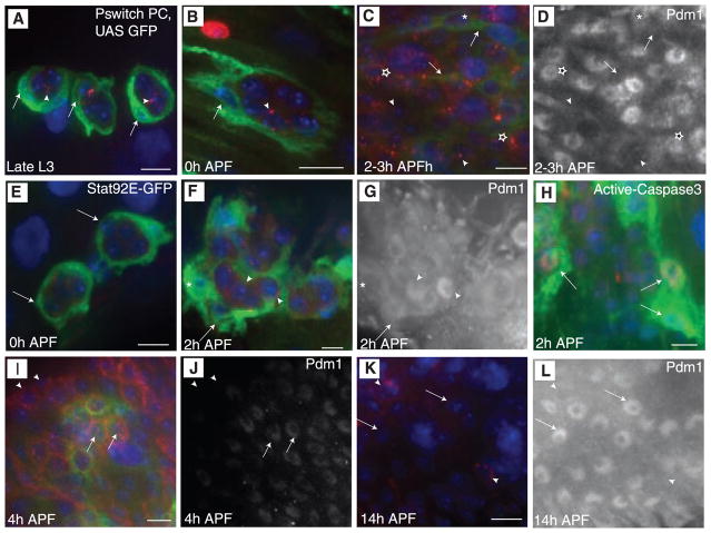
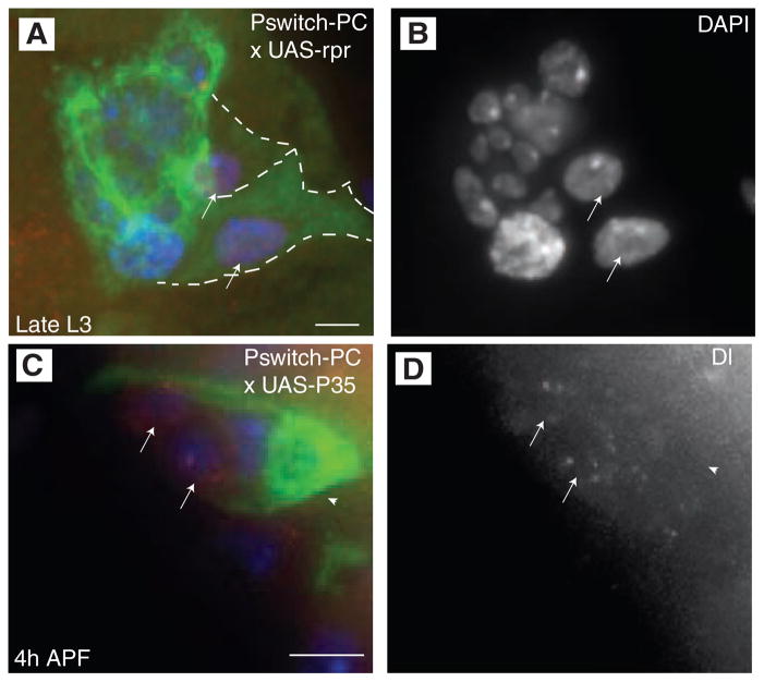
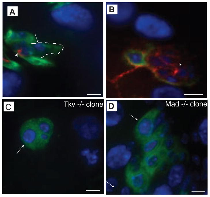
Stem cell niches are locations where stem cells reside and self-renew. Although studies have shown how niches maintain stem cell fate during tissue homeostasis, less is known about their roles in establishing stem cells. The adult Drosophila midgut is maintained by intestinal stem cells (ISCs); however, how they are established is unknown. Here, we show that an ISC progenitor generates a niche cell via Notch signaling. This niche uses the bone morphogenetic protein 2/4 homolog, decapentaplegic, to allow progenitors to divide in an undifferentiated state and subsequently breaks down and dies, resulting in the specification of ISCs in the adult midgut. Our results demonstrate a paradigm for stem cell-niche biology, where progenitors generate transient niches that determine stem cell fate and may give insights into stem cell specification in other tissues.
Figures
Similar articles
-
Sara endosomes and the asymmetric division of intestinal stem cells.Development. 2014 May;141(10):2014-23. doi: 10.1242/dev.104240. Development. 2014. PMID: 24803650
-
Intestinal epithelium-derived BMP controls stem cell self-renewal in Drosophila adult midgut.Elife. 2014 Mar 11;3:e01857. doi: 10.7554/eLife.01857. Elife. 2014. PMID: 24618900 Free PMC article.
-
Origin and dynamic lineage characteristics of the developing Drosophila midgut stem cells.Dev Biol. 2016 Aug 15;416(2):347-60. doi: 10.1016/j.ydbio.2016.06.018. Epub 2016 Jun 16. Dev Biol. 2016. PMID: 27321560
-
Intestinal stem cells in the adult Drosophila midgut.Exp Cell Res. 2011 Nov 15;317(19):2780-8. doi: 10.1016/j.yexcr.2011.07.020. Epub 2011 Aug 11. Exp Cell Res. 2011. PMID: 21856297 Free PMC article. Review.
-
Another notch in stem cell biology: Drosophila intestinal stem cells and the specification of cell fates.Bioessays. 2008 Feb;30(2):107-9. doi: 10.1002/bies.20710. Bioessays. 2008. PMID: 18200564 Review.
Cited by
-
α-Phenylalanyl tRNA synthetase competes with Notch signaling through its N-terminal domain.PLoS Genet. 2022 Apr 29;18(4):e1010185. doi: 10.1371/journal.pgen.1010185. eCollection 2022 Apr. PLoS Genet. 2022. PMID: 35486661 Free PMC article.
-
lines and bowl affect the specification of cyst stem cells and niche cells in the Drosophila testis.Development. 2011 May;138(9):1687-96. doi: 10.1242/dev.057364. Development. 2011. PMID: 21486923 Free PMC article.
-
Defining and redefining the nephron progenitor population.Pediatr Nephrol. 2011 Sep;26(9):1395-406. doi: 10.1007/s00467-010-1750-4. Epub 2011 Jan 14. Pediatr Nephrol. 2011. PMID: 21229268 Free PMC article. Review.
-
Cell Competition Modifies Adult Stem Cell and Tissue Population Dynamics in a JAK-STAT-Dependent Manner.Dev Cell. 2015 Aug 10;34(3):297-309. doi: 10.1016/j.devcel.2015.06.010. Epub 2015 Jul 23. Dev Cell. 2015. PMID: 26212135 Free PMC article.
-
Signal Integration by the IκB Protein Pickle Shapes Drosophila Innate Host Defense.Cell Host Microbe. 2016 Sep 14;20(3):283-295. doi: 10.1016/j.chom.2016.08.003. Cell Host Microbe. 2016. PMID: 27631699 Free PMC article.
References
Publication types
MeSH terms
Substances
Grants and funding
LinkOut - more resources
Full Text Sources
Other Literature Sources
Molecular Biology Databases
