. 2010 Mar;298(3):G467-73.
doi: 10.1152/ajpgi.00360.2009. Epub 2010 Jan 7.

Green tea catechin EGCG inhibits ileal apical sodium bile acid transporter ASBT

Affiliations

Green tea catechin EGCG inhibits ileal apical sodium bile acid transporter ASBT

Fadi Annaba et al. Am J Physiol Gastrointest Liver Physiol. 2010 Mar.

Abstract

Green tea catechins exhibit hypocholesterolemic effects probably via their inhibitory effects on intestinal bile acid absorption. Ileal apical sodium-dependent bile acid transporter (ASBT) is responsible for reabsorption of bile acids. The present studies were, therefore, designed to investigate the modulation of ASBT function and membrane expression by green tea catechins in human embryonic kidney HEK-293 cells stably transfected with ASBT-V5 fusion protein and intestinal Caco-2 monolayers. Our data showed that ASBT activity was significantly decreased by (-)-epigallocatechin-3-gallate (EGCG) but not other green tea catechins. Inhibition of PKC, phosphatidylinositol 3-kinase, and MAPK-dependent pathways failed to block the reduction in ASBT activity by EGCG. Kinetics studies showed a significant decrease in the V(max) of the transporter, whereas total ASBT content on the plasma membrane was unaltered by EGCG. Concomitant with the decrease in ASBT function, EGCG significantly reduced ASBT pool in the detergent-insoluble fraction, while increasing its presence in the detergent-soluble fraction of plasma membrane. Furthermore, EGCG decreased the association of ASBT with floating lipid raft fractions of cellular membrane on Optiprep density gradient. In conclusion, our data demonstrate a novel role of lipid rafts in the modulation of ASBT function by the dietary component EGCG, which may underlie the hypocholesterolemic effects of green tea.


Figures
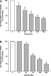
Fig. 1.
Fig. 1.
ASBT is inhibited by (−)-epigallocatechin-3-gallate (EGCG). A: 2BT cells were incubated with 50 μM of EGCG for different periods of time. Cells were then washed with tracer-free uptake buffer and subsequently incubated with the uptake buffer containing 10 μM of [3H]taurocholic acid (TC). Uptake values were determined as described in material and methods and expressed as % of control (untreated cells). B: 2BT cells were incubated for 90 min with different concentrations of EGCG. TC uptake was then assessed as described in material and methods and values were expressed as % of control (untreated cells). Values in A and B are expressed as means ± SE of 6–9 determinations performed on 3 separate occasions. *P ≤ 0.05 compared with control.
Fig. 2.
Fig. 2.
EGCG-induced inhibition of ASBT is specific. 2BT cells were incubated for 90 min with 50 μM of EGCG, (−)-catechin (C), (−)-catechin gallate (CG), (−)-epicatechin (EC), (−)-epicatechin gallate (ECG), or (−)-epigallocatechin (EGC). TC uptake was evaluated as described in material and methods and values were expressed as % of control (untreated cells). Results are presented as means ± SE of at least 6 determinations from 3 separate occasions. *P ≤ 0.05 compared with control.
Fig. 3.
Fig. 3.
Cellular signaling pathways are not involved in ASBT inhibition by EGCG. 2BT cells were preincubated for 30 min with 5 μM bisindolylmaleimide I HCl (BIM), 50 μM LY294002, 5 μg/ml dephostatin, or 10 μM of PD98095. Cells were then incubated with each inhibitor along with 50 μM EGCG for additional 90 min. Uptake was performed as described in material and methods and values were expressed as % of control (cells without EGCG treatment). Values are shown as means ± SE of at least 9 determinations from 3–4 separate occasions. *P ≤ 0.05 compared with control.
Fig. 4.
Fig. 4.
EGCG decreases the Vmax of ASBT. 2BT cells were incubated for 90 min with 50 μM EGCG and then TC uptake was performed in the presence of increasing concentrations of the substrate TC. Kinetic parameters were obtained by the Michaelis-Menten analysis utilizing GraphPad prism software. A representative experiment is shown in the figure, and uptake values are presented as pmol·mg protein−1·5 min−1.
Fig. 5.
Fig. 5.
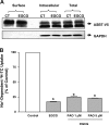
Inhibition by EGCG is not mediated by alterations in ASBT level on plasma membrane. A: EGCG (50 μM) was added to 2BT cells for 90 min, and cell surface biotinylation was then performed as described in material and methods. Proteins from total lysates, intracellular fractions, and biotinylated fractions (surface fractions) were then separated on 10% SDS-PAGE gel and electrotransferred to nitrocellulose blots. Blots were then probed with anti-V5 antibodies and bands corresponding to ASBT-V5 fusion proteins were visualized as described in material and methods. ASBT-V5 fusion protein appears as multiple bands representing glycosylated and unglycosylated polypeptides. CT, control. B: 2BT cells were preincubated for 30 min with 1 or 5 μM of phenylarsine oxide (PAO) alone and then with 50 μM of EGCG for additional 90 min. TC uptake was then performed as described in material and methods. Values represent means ± SE of 6–9 determinations performed on 3 separate occasions. *P ≤ 0.05 compared with control (cells that were not treated with EGCG).
Fig. 6.
Fig. 6.
EGCG reduces ASBT partition in the detergent insoluble (DI) fraction of cellular membranes. 2BT cells were incubated with 50 μM of EGCG or EC for 90 min and then washed and lysed in buffer without a detergent as material and methods. In a separate group of cells, EGCG was washed out and cells were left for additional 24 h after EGCG treatment before lysing (washout). Cellular membranes were then isolated by ultracentrifugation and then incubated for 30 min at 4°C in a buffer containing 1% Triton X-100. Detergent soluble (DS) and DI fractions were then isolated as described in material and methods. Proteins were separated on 10% SDS-PAGE gel and blots were probed with anti-V5 antibodies to visualize the ASBT-V5 fusion proteins.
Fig. 7.
Fig. 7.
Inhibition by EGCG is reversible. EGCG (50 μM) was added to 2BT cells for 90 min then cells were thoroughly washed and left for additional 24 h and then TC uptake was evaluated (washout cells). A second group of cells were treated with EGCG for 90 min and TC uptake was then performed at the end of the incubation as described in the legend for Fig. 1. Uptake values are presented as % of control and expressed as means ± SE of 6–9 determinations performed on 3 separate occasions. *P ≤ 0.05 compared with control (untreated cells).
Fig. 8.
Fig. 8.
EGCG causes redistribution of ASBT to the high-density fractions on density gradient. 2BT cells were incubated with 50 μM of EGCG for 90 min and then washed and lysed in homogenization buffer without a detergent as described in material and methods. Cellular membranes were then isolated by ultracentrifugation and then incubated in TNE buffer as described in material and methods. The membranes were then laid at the bottom of Optiprep density gradient and subjected to ultracentrifugation. Fractions were then collected from the top of the gradient (low-density fractions) to the bottom of the gradient (high-density fractions). Proteins in the fractions were separated on 10% SDS-PAGE and blots were probed with anti-V5 or anti-flotillin antibodies.
Fig. 9.
Fig. 9.
EGCG inhibits ASBT function in the human intestinal cell line Caco-2. Postconfluent Caco-2 monolayers plated on plastic support in 24-well plates and incubated for 90 min with different concentrations of EGCG. Cells were then washed and TC uptake was assessed as described in material and methods. Uptake values are expressed as % of control and presented as means ± SE from 3–6 determinations performed on 3 separate occasions. *P ≤ 0.05 compared with control (untreated cells).

Similar articles

Cited by

References

    1. Adachi S, Nagao T, Ingolfsson HI, Maxfield FR, Andersen OS, Kopelovich L, Weinstein IB. The inhibitory effect of (−)-epigallocatechin gallate on activation of the epidermal growth factor receptor is associated with altered lipid order in HT29 colon cancer cells. Cancer Res 67: 6493–6501, 2007 - PubMed
    1. Albrecht DS, Clubbs EA, Ferruzzi M, Bomser JA. Epigallocatechin-3-gallate (EGCG) inhibits PC-3 prostate cancer cell proliferation via MEK-independent ERK1/2 activation. Chem Biol Interact 171: 89–95, 2008 - PubMed
    1. Alpini G, Glaser S, Baiocchi L, Francis H, Xia X, Lesage G. Secretin activation of the apical Na+-dependent bile acid transporter is associated with cholehepatic shunting in rats. Hepatology 41: 1037–1045, 2005 - PubMed
    1. Alrefai WA, Gill RK. Bile acid transporters: structure, function, regulation and pathophysiological implications. Pharm Res 24: 1803–1823, 2007 - PubMed
    1. Alrefai WA, Sarwar Z, Tyagi S, Saksena S, Dudeja PK, Gill RK. Cholesterol modulates human intestinal sodium-dependent bile acid transporter. Am J Physiol Gastrointest Liver Physiol 288: G978–G985, 2005 - PubMed

Publication types

MeSH terms

LinkOut - more resources