

Reduced sox9 function promotes heart valve calcification phenotypes in vivo
- PMID: 20056916
- PMCID: PMC2863131
- DOI: 10.1161/CIRCRESAHA.109.213702
Reduced sox9 function promotes heart valve calcification phenotypes in vivo
Abstract
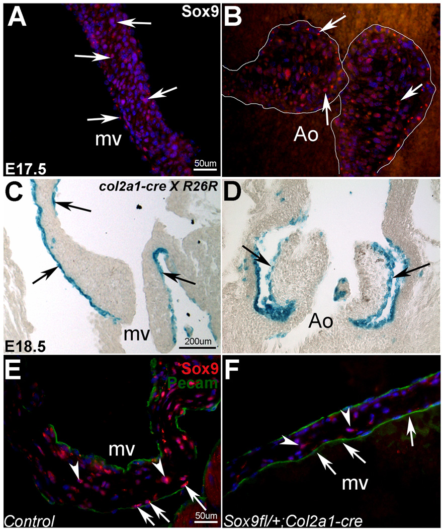
Rationale: Calcification of heart valve structures is the most common form of valvular disease and is characterized by the appearance of bone-like phenotypes within affected structures. Despite the clinical significance, the underlying etiology of disease onset and progression is largely unknown and valve replacement remains the most effective treatment. The SRY-related transcription factor Sox9 is expressed in developing and mature heart valves, and its function is required for expression of cartilage-associated proteins, similar to its role in chondrogenesis. In addition to cartilage-associated defects, mice with reduced sox9 function develop skeletal bone prematurely; however, the ability of sox9 deficiency to promote ectopic osteogenic phenotypes in heart valves has not been examined.
Objective: This study aims to determine the role of Sox9 in maintaining connective tissue homeostasis in mature heart valves using in vivo and in vitro approaches.
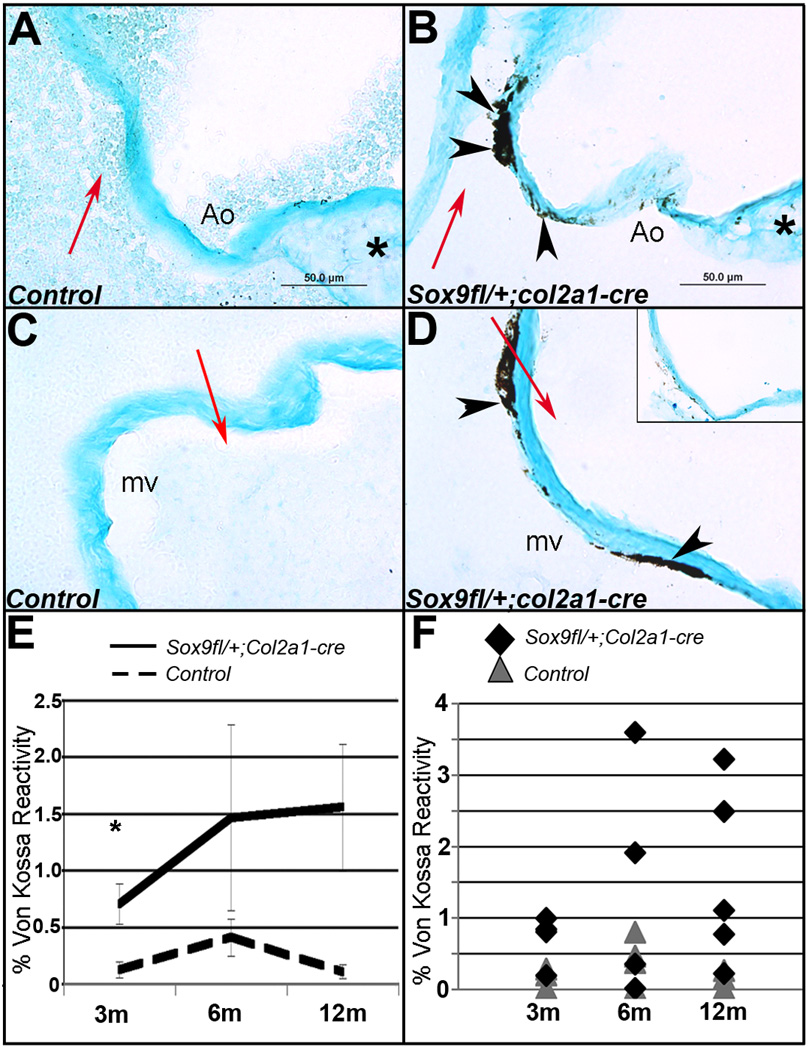
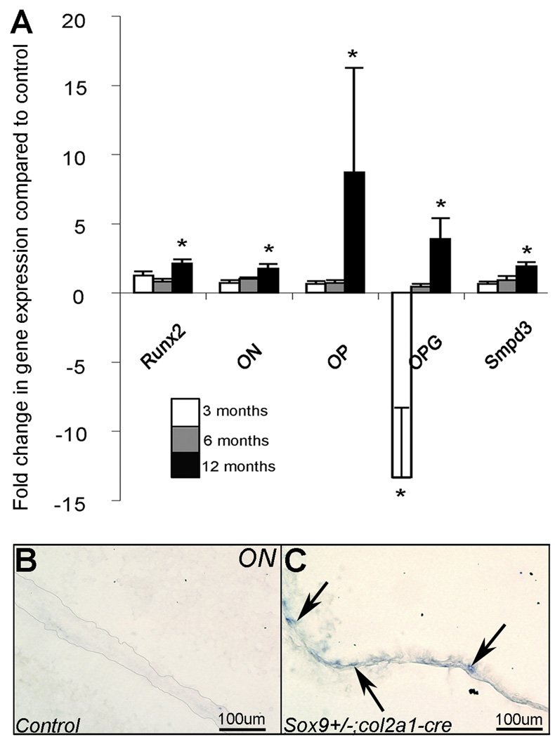
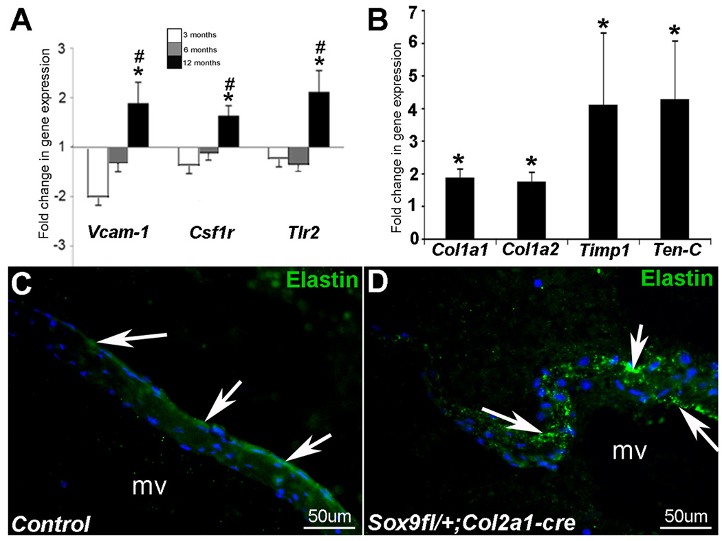
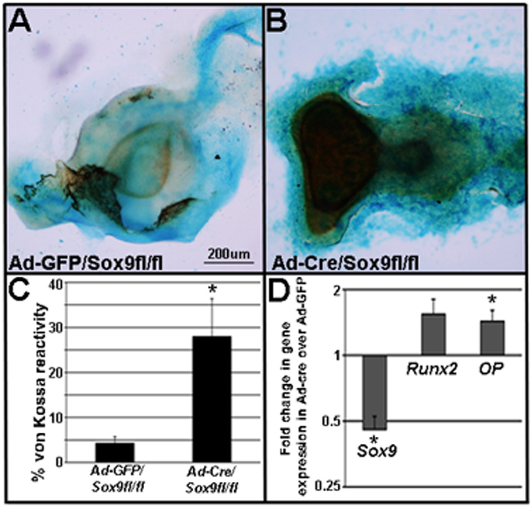
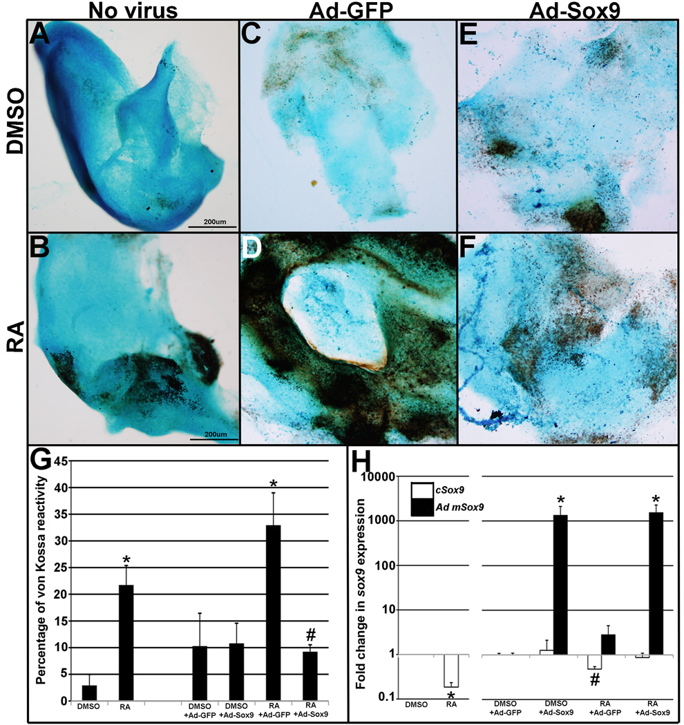
Methods and results: Using histological and molecular analyses, we report that, from 3 months of age, Sox9(fl/+);Col2a1-cre mice develop calcific lesions in heart valve leaflets associated with increased expression of bone-related genes and activation of inflammation and matrix remodeling processes. Consistently, ectopic calcification is also observed following direct knockdown of Sox9 in heart valves in vitro. Furthermore, we show that retinoic acid treatment in mature heart valves is sufficient to promote calcific processes in vitro, which can be attenuated by Sox9 overexpression.
Conclusions: This study provides insight into the molecular mechanisms of heart valve calcification and identifies reduced Sox9 function as a potential genetic basis for calcific valvular disease.
Figures
Similar articles
-
Cadherin-11 Overexpression Induces Extracellular Matrix Remodeling and Calcification in Mature Aortic Valves.Arterioscler Thromb Vasc Biol. 2016 Aug;36(8):1627-37. doi: 10.1161/ATVBAHA.116.307812. Epub 2016 Jun 16. Arterioscler Thromb Vasc Biol. 2016. PMID: 27312222 Free PMC article.
-
Increased dietary intake of vitamin A promotes aortic valve calcification in vivo.Arterioscler Thromb Vasc Biol. 2013 Feb;33(2):285-93. doi: 10.1161/ATVBAHA.112.300388. Epub 2012 Nov 29. Arterioscler Thromb Vasc Biol. 2013. PMID: 23202364 Free PMC article.
-
Valve Endothelial Cell-Derived Tgfβ1 Signaling Promotes Nuclear Localization of Sox9 in Interstitial Cells Associated With Attenuated Calcification.Arterioscler Thromb Vasc Biol. 2016 Feb;36(2):328-38. doi: 10.1161/ATVBAHA.115.306091. Epub 2015 Dec 3. Arterioscler Thromb Vasc Biol. 2016. PMID: 26634652 Free PMC article.
-
Mechanisms of mitral annular calcification.Trends Cardiovasc Med. 2020 Jul;30(5):289-295. doi: 10.1016/j.tcm.2019.07.011. Epub 2019 Aug 5. Trends Cardiovasc Med. 2020. PMID: 31402089 Review.
-
Conserved transcriptional regulatory mechanisms in aortic valve development and disease.Arterioscler Thromb Vasc Biol. 2014 Apr;34(4):737-41. doi: 10.1161/ATVBAHA.113.302071. Arterioscler Thromb Vasc Biol. 2014. PMID: 24665126 Free PMC article. Review.
Cited by
-
Ferritin in Kidney and Vascular Related Diseases: Novel Roles for an Old Player.Pharmaceuticals (Basel). 2019 Jun 21;12(2):96. doi: 10.3390/ph12020096. Pharmaceuticals (Basel). 2019. PMID: 31234273 Free PMC article. Review.
-
Differential activation of valvulogenic, chondrogenic, and osteogenic pathways in mouse models of myxomatous and calcific aortic valve disease.J Mol Cell Cardiol. 2012 Mar;52(3):689-700. doi: 10.1016/j.yjmcc.2011.12.013. Epub 2012 Jan 10. J Mol Cell Cardiol. 2012. PMID: 22248532 Free PMC article.
-
Angiopoietin-like 2 is essential to aortic valve development in mice.Commun Biol. 2022 Nov 21;5(1):1277. doi: 10.1038/s42003-022-04243-6. Commun Biol. 2022. PMID: 36414704 Free PMC article.
-
Cadherin-11 Overexpression Induces Extracellular Matrix Remodeling and Calcification in Mature Aortic Valves.Arterioscler Thromb Vasc Biol. 2016 Aug;36(8):1627-37. doi: 10.1161/ATVBAHA.116.307812. Epub 2016 Jun 16. Arterioscler Thromb Vasc Biol. 2016. PMID: 27312222 Free PMC article.
-
The Genetic Regulation of Aortic Valve Development and Calcific Disease.Front Cardiovasc Med. 2018 Nov 6;5:162. doi: 10.3389/fcvm.2018.00162. eCollection 2018. Front Cardiovasc Med. 2018. PMID: 30460247 Free PMC article. Review.
References
-
- Stewart B, Siscovick D, Lind B, Gardin J, Gottdiener J, Smith V, Kitzman D, Otto C. Clinical factors associated with calcific aortic valvular disease. J Am Coll Cardiol. 1997;29:630–634. - PubMed
-
- Otto CM. Calcific aortic stenosis - time to look more closely at the valve. N Engl J Med. 2008;359:1395–1398. - PubMed
-
- Hinton RB, Jr, Lincoln J, Deutsch GH, Osinska H, Manning PB, Benson DW, Yutzey KE. Extracellular matrix remodeling and organization in developing and diseased aortic valves. Circ Res. 2006;98:1431–1438. - PubMed
-
- Lincoln J, Lange AW, Yutzey KE. Hearts and bones: Shared regulatory mechanisms in heart valve, cartilage, tendon, and bone development. Developmental Biology. 2006;294:292–302. - PubMed
-
- Rabkin E, Aikawa M, Stone JR, Fukumoto Y, Libby P, Schoen FJ. Activated interstitial myofibroblasts express catabolic enzymes and mediate matrix remodeling in myxomatous heart valves. Circulation. 2001;104:2525–2532. - PubMed
Publication types
MeSH terms
Substances
Grants and funding
LinkOut - more resources
Full Text Sources
Other Literature Sources
Medical
Molecular Biology Databases
Research Materials
