

Review
doi: 10.1161/CIRCRESAHA.109.207456.
Intramyocardial fibroblast myocyte communication
Affiliations
- PMID: 20056945
- PMCID: PMC2805465
- DOI: 10.1161/CIRCRESAHA.109.207456
Item in Clipboard
Review
Intramyocardial fibroblast myocyte communication
Circ Res.
.
Abstract
Cardiac fibroblasts are emerging as key components of normal cardiac function, as well as the response to stressors and injury. These most numerous cells of the heart interact with myocytes via paracrine mechanisms, alterations in extracellular matrix homeostasis, and direct cell-cell interactions. It is possible that they are a contributor to the inability of adult myocytes to proliferate and may influence cardiac progenitor biology. Furthering our understanding of how cardiac fibroblasts and myocytes interact may provide an avenue to novel treatments for heart failure prevention. This review discusses the most recent concepts in cardiac fibroblast-myocyte communication and areas of potential future research.
Figures
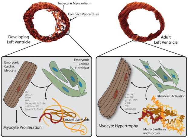
In the developing ventricle, embryonic cardiac fibroblasts reside within the outer, compacting myocardium. These fibroblasts express components of the extracellular matrix, including fibronectin and collagen, as well as heparin-binding EGF-like growth factor, that may promote myocyte proliferation.(8) Intracellular signals are activated by myocyte integration of fibroblast-derived paracrine stimuli and signals from matrix proteins to effect coordinated proliferation and organogenesis. In contrast, under biomechanical stress, adult cardiac myocytes respond in a hypertrophic rather than proliferative manner, in part fostered by paracrine signals from neighboring cardiac fibroblasts and interactions with the remodeling extracellular matrix. Again, numerous intracellular signals systems are activated to effect a coordinated hypertrophic program.(163)
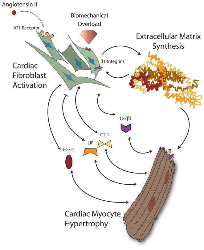
Under biomechanical overload, cardiac fibroblasts and myocytes respond to an altered environment via multiple mechanisms including integrin-extracellular matrix interactions and renin-angiotensin-aldosterone axis activation. Cardiac fibroblasts increase synthesis of matrix proteins and secrete a variety of paracrine factors that can stimulate myocyte hypertrophy. Cardiac myocytes similarly respond by secreting a conglomerate of factors. Hormones such as TGFβ1, FGF-2, and the IL-6 family members LIF and CT-1 have all been implicated in this bidirectional fibroblast-myocyte hormonal crosstalk.
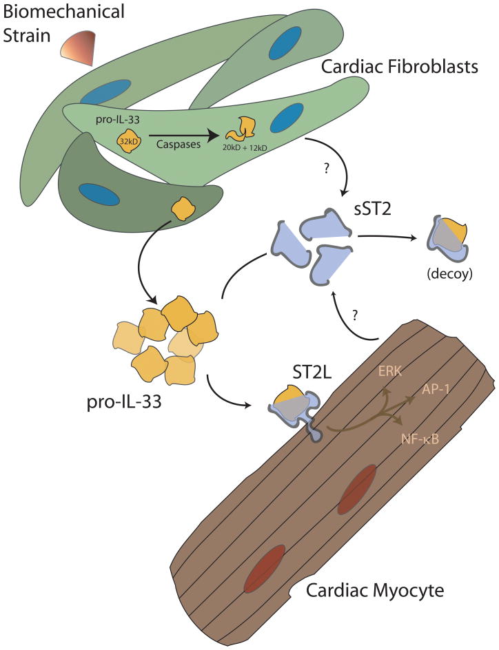
The IL-33/ST2 system is a recently-described cardiac fibroblast-myocyte signaling system. This signaling pathway serves an anti-hypertrophic and cardioprotective mechanism in the face of biomechanical overload. IL-33 is produced by mechanically-loaded cardiac fibroblasts and is possibly inactivated by caspases. Extracellular IL-33 may bind to a soluble “decoy” form of its receptor, sST2, and be removed from the biologically available pool, or bind a transmembrane form, ST2L, on the surface of cardiac myocytes. In the face of pro-hypertrophic stimuli in vitro or pressure overload in vivo, IL-33 appears to confer anti-hypertrophic and anti-fibrotic properties to the myocardium.(93, 97)
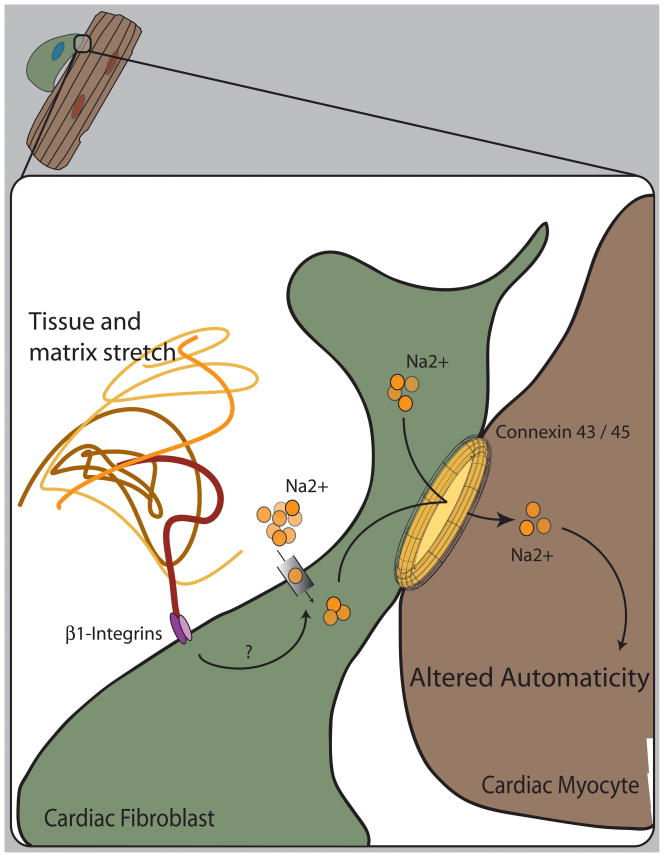
Both electrophysiologic and molecular data suggest that cardiac fibroblasts and myocytes are in direct contact and part of a syncytial network afforded by the conglomerate of connexin 43 and 45 hemichannels. Mechanical stretch alters the electrical state of cardiac fibroblasts by activation of a combination of ion selective and non-selective channels.(146, 147) Fibroblasts may acquire and affect the synchronized beating of myocytes in culture. Because fibroblasts are abundant within the region of the sinoatrial node of the mammalian heart, it is plausible that tissue strain, mediated by integrin signaling, affects cardiac fibroblast depolarization and possible intracellular kinase signaling to transmit signals to neighboring cardiac myocytes, altering rhythmic automaticity.
References
-
- Brilla CG, Maisch B, Weber KT. Myocardial collagen matrix remodelling in arterial hypertension. Eur Heart J. 1992;13(Suppl D):24–32. - PubMed
-
- Weber KT, Brilla CG. Myocardial fibrosis and the renin-angiotensin-aldosterone system. J Cardiovasc Pharmacol. 1992;20(Suppl 1):S48–54. - PubMed
-
- Weber KT, Brilla CG, Campbell SE, Zhou G, Matsubara L, Guarda E. Pathologic hypertrophy with fibrosis: the structural basis for myocardial failure. Blood Press. 1992;1:75–85. - PubMed
-
- Weber KT, Brilla CG, Janicki JS, Reddy HK, Campbell SE. Myocardial fibrosis: role of ventricular systolic pressure, arterial hypertension, and circulating hormones. Basic Res Cardiol. 1991;86(Suppl 3):25–31. - PubMed
-
- Weber KT, Jalil JE, Janicki JS, Pick R. Myocardial collagen remodeling in pressure overload hypertrophy. A case for interstitial heart disease. Am J Hypertens. 1989;2:931–940. - PubMed
Publication types
MeSH terms
Grants and funding
LinkOut - more resources
Full Text Sources
Other Literature Sources
Medical
