Cloning, expression and crystallization of dihydrodipicolinate reductase from methicillin-resistant Staphylococcus aureus
- PMID: 20057072
- PMCID: PMC2805538
- DOI: 10.1107/S1744309109047964
Cloning, expression and crystallization of dihydrodipicolinate reductase from methicillin-resistant Staphylococcus aureus
Abstract
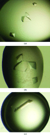
Dihydrodipicolinate reductase (DHDPR; EC 1.3.1.26) catalyzes the nucleotide (NADH/NADPH) dependent second step of the lysine-biosynthesis pathway in bacteria and plants. Here, the cloning, expression, purification, crystallization and preliminary X-ray diffraction analysis of DHDPR from methicillin-resistant Staphylococcus aureus (MRSA-DHDPR) are presented. The enzyme was crystallized in a number of forms, predominantly with ammonium sulfate as a precipitant, with the best crystal form diffracting to beyond 3.65 A resolution. Crystal structures of the apo form as well as of cofactor (NADPH) bound and inhibitor (2,6-pyridinedicarboxylate) bound forms of MRSA-DHDPR will provide insight into the structure and function of this essential enzyme and valid drug target.
Figures


Similar articles
-
Comparative structure and function analyses of native and his-tagged forms of dihydrodipicolinate reductase from methicillin-resistant Staphylococcus aureus.Protein Expr Purif. 2012 Sep;85(1):66-76. doi: 10.1016/j.pep.2012.06.017. Epub 2012 Jul 6. Protein Expr Purif. 2012. PMID: 22776412
-
Catalytic mechanism and cofactor preference of dihydrodipicolinate reductase from methicillin-resistant Staphylococcus aureus.Arch Biochem Biophys. 2011 Aug 15;512(2):167-74. doi: 10.1016/j.abb.2011.06.006. Epub 2011 Jun 16. Arch Biochem Biophys. 2011. PMID: 21704017
-
The structure of dihydrodipicolinate reductase (DapB) from Mycobacterium tuberculosis in three crystal forms.Acta Crystallogr D Biol Crystallogr. 2010 Jan;66(Pt 1):61-72. doi: 10.1107/S0907444909043960. Epub 2009 Dec 21. Acta Crystallogr D Biol Crystallogr. 2010. PMID: 20057050
-
Structure and nucleotide specificity of Staphylococcus aureus dihydrodipicolinate reductase (DapB).FEBS Lett. 2011 Aug 19;585(16):2561-7. doi: 10.1016/j.febslet.2011.07.021. Epub 2011 Jul 26. FEBS Lett. 2011. PMID: 21803042
-
Cloning, expression, purification, crystallization and preliminary X-ray diffraction analysis of DapB (Rv2773c) from Mycobacterium tuberculosis.Acta Crystallogr Sect F Struct Biol Cryst Commun. 2005 Jul 1;61(Pt 7):718-21. doi: 10.1107/S174430910501938X. Epub 2005 Jun 30. Acta Crystallogr Sect F Struct Biol Cryst Commun. 2005. PMID: 16511139 Free PMC article.
Cited by
-
Characterisation of the first enzymes committed to lysine biosynthesis in Arabidopsis thaliana.PLoS One. 2012;7(7):e40318. doi: 10.1371/journal.pone.0040318. Epub 2012 Jul 5. PLoS One. 2012. PMID: 22792278 Free PMC article.
-
Molecular evolution of an oligomeric biocatalyst functioning in lysine biosynthesis.Biophys Rev. 2018 Apr;10(2):153-162. doi: 10.1007/s12551-017-0350-y. Epub 2017 Dec 5. Biophys Rev. 2018. PMID: 29204887 Free PMC article. Review.
-
Cloning, expression, purification and crystallization of dihydrodipicolinate synthase from the grapevine Vitis vinifera.Acta Crystallogr Sect F Struct Biol Cryst Commun. 2011 Dec 1;67(Pt 12):1537-41. doi: 10.1107/S1744309111038395. Epub 2011 Nov 25. Acta Crystallogr Sect F Struct Biol Cryst Commun. 2011. PMID: 22139160 Free PMC article.
-
Cloning, expression, purification and crystallization of dihydrodipicolinate synthase from Agrobacterium tumefaciens.Acta Crystallogr Sect F Struct Biol Cryst Commun. 2012 Sep 1;68(Pt 9):1040-7. doi: 10.1107/S1744309112033052. Epub 2012 Aug 30. Acta Crystallogr Sect F Struct Biol Cryst Commun. 2012. PMID: 22949190 Free PMC article.
-
Structure and Function of Cyanobacterial DHDPS and DHDPR.Sci Rep. 2016 Nov 15;6:37111. doi: 10.1038/srep37111. Sci Rep. 2016. PMID: 27845445 Free PMC article.
References
Publication types
MeSH terms
Substances
LinkOut - more resources
Full Text Sources
Medical

