

doi: 10.1107/S1744309109050593.
Epub 2009 Dec 25.
Crystallization and preliminary crystallographic analysis of the measles virus hemagglutinin in complex with the CD46 receptor
Affiliations
- PMID: 20057080
- PMCID: PMC2805546
- DOI: 10.1107/S1744309109050593
Item in Clipboard
Crystallization and preliminary crystallographic analysis of the measles virus hemagglutinin in complex with the CD46 receptor
Acta Crystallogr Sect F Struct Biol Cryst Commun.
.
Abstract
The measles virus (MV) hemagglutinin (MV-H) mediates the attachment of MV particles to cell-surface receptors for entry into host cells. MV uses two receptors for attachment to host cells: the complement-control protein CD46 and the signalling lymphocyte activation molecule (SLAM). The MV-H glycoprotein from an Edmonston MV variant and the MV-binding fragment of the CD46 receptor were overproduced in mammalian cells and used to crystallize an MV-H-CD46 complex. Well diffracting crystals containing two complexes in the asymmetric unit were obtained and the structure of the complex was solved by the molecular-replacement method.
Figures
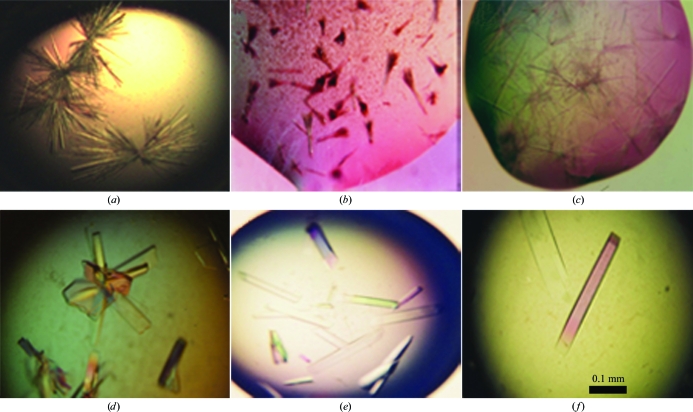
Crystals of the isolated MV-H fragment and the MV-H–CD46 complex. The top row shows crystals prepared with 2 M ammonium sulfate, 5% MPD and 0.1 M sodium cadodylate pH 6.5. The bottom row shows crystals prepared with 12% PEG 8000, 0.2 M ammonium sulfate, 2% PEG 400 and 0.1 M sodium MES pH 6.5. (a) Crystals of the MV-H fragment. (b) Crystals of endoglycosidase H-treated MV-H fragment. (c, d) Crystals of the MV-H–CD46 complex. (e) Crystals of the complex prepared with the addition of 1% 1,2,3-heptanetriol to the PEG 8000 crystallization condition. (f) Crystals of the complex prepared with the addition of 1% 1,2,3-heptanetriol and 1 mM sodium tungstate (Na2WO4).
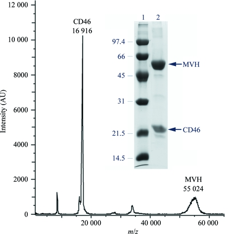
Identification of the proteins in the MV-H–CD46 crystals. MALDI–TOF analysis of redissolved crystals. The spectrum shows two peaks that correspond to proteins with molecular masses of 55 024 and 16 916 Da, corresponding to MV-H and CD46, respectively. Mass determination was calculated from the single charged ion of the proteins. The inset shows 12% SDS–PAGE analysis under reducing conditions of redissolved crystals (lane 2) and molecular mass markers (lane 1). The mass (kDa) and migration of the marker proteins are indicated.
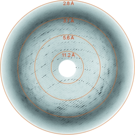
X-ray diffraction image from a native MV-H–CD46 crystal (Na7 in Table 1 ▶). Rings are shown at the detector edge and at three additional resolution limits.
References
-
- Brünger, A. T., Adams, P. D., Clore, G. M., DeLano, W. L., Gros, P., Grosse-Kunstleve, R. W., Jiang, J.-S., Kuszewski, J., Nilges, M., Pannu, N. S., Read, R. J., Rice, L. M., Simonson, T. & Warren, G. L. (1998). Acta Cryst. D54, 905–921. - PubMed
-
- Casasnovas, J. M. & Springer, T. A. (1995). J. Biol. Chem.270, 13216–13224. - PubMed
-
- Colf, L. A., Juo, Z. S. & Garcia, K. C. (2007). Nature Struct. Mol. Biol.14, 1227–1228. - PubMed
-
- Dorig, R. E., Marcil, A., Chopra, A. & Richardson, C. D. (1993). Cell, 75, 295–305. - PubMed
Publication types
MeSH terms
Substances
Grants and funding
LinkOut - more resources
Full Text Sources
