. 2010 Feb 17;29(4):819-29.
doi: 10.1038/emboj.2009.394. Epub 2010 Jan 7.

HipHop interacts with HOAP and HP1 to protect Drosophila telomeres in a sequence-independent manner

Affiliations

HipHop interacts with HOAP and HP1 to protect Drosophila telomeres in a sequence-independent manner

Guanjun Gao et al. EMBO J. .

Abstract

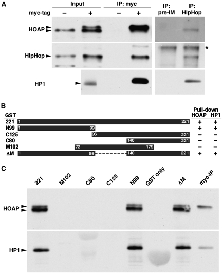
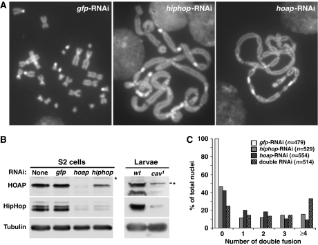
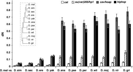
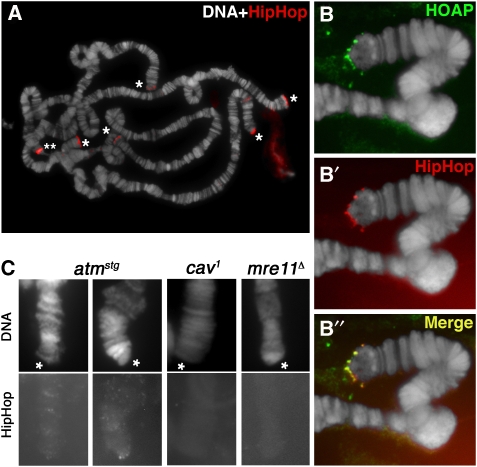
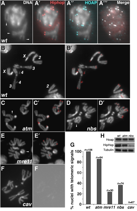
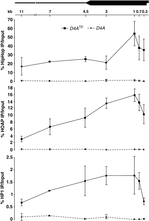
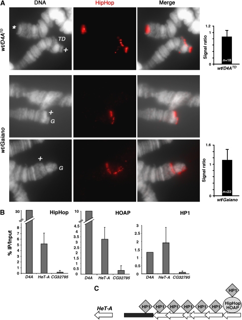
Telomeres prevent chromosome ends from being repaired as double-strand breaks (DSBs). Telomere identity in Drosophila is determined epigenetically with no sequence either necessary or sufficient. To better understand this sequence-independent capping mechanism, we isolated proteins that interact with the HP1/ORC-associated protein (HOAP) capping protein, and identified HipHop as a subunit of the complex. Loss of one protein destabilizes the other and renders telomeres susceptible to fusion. Both HipHop and HOAP are enriched at telomeres, where they also interact with the conserved HP1 protein. We developed a model telomere lacking repetitive sequences to study the distribution of HipHop, HOAP and HP1 using chromatin immunoprecipitation (ChIP). We discovered that they occupy a broad region >10 kb from the chromosome end and their binding is independent of the underlying DNA sequence. HipHop and HOAP are both rapidly evolving proteins yet their telomeric deposition is under the control of the conserved ATM and Mre11-Rad50-Nbs (MRN) proteins that modulate DNA structures at telomeres and at DSBs. Our characterization of HipHop and HOAP reveals functional analogies between the Drosophila proteins and subunits of the yeast and mammalian capping complexes, implicating conservation in epigenetic capping mechanisms.


Conflict of interest statement

The authors declare that they have no conflict of interest.

Figures

Figure 1
Figure 1
HipHop interacts with HOAP and HP1. (A) Reciprocal IP between HipHop and HOAP. In the left three panels, embryonic extracts with (+) or without (−) myc-HOAP were subjected to anti-myc IP, followed by western blot analyses with antibodies against the antigens indicated to the left. Input samples were loaded as controls. The arrowheads mark the position of the proteins of interest. For HOAP, the two arrowheads mark myc-HOAP (upper) and HOAP (lower), respectively, as the extracts were from embryos having both myc-cav and cav alleles (see Supplementary data for explanation). In the right three panels, wild-type (untagged) extracts were subjected to IP with either a HipHop antibody (HipHop) or its corresponding pre-immune serum (pre-IM). In the HipHop western blot, a non-specific band is marked with *, which also served as a loading control. (B) GST-HipHop fusion proteins used to map the HOAP-interacting domain. The names of the construct are listed to the left of the black boxes, which indicate the regions of HipHop protein fused with GST. The right table summarizes the pull-down results (shown in C), with ‘+' indicating a positive interaction, and ‘−' for a lack of interaction. (C) GST-HipHop pull-down results. The names of the fusion constructs are listed on top of the membranes. The panels show GST pull down using embryonic extracts with both myc-HOAP and HOAP proteins, followed by western blot probed with antibodies listed to the left. Arrowheads mark the proteins of interest. In the last lane, a sample from anti-myc IP (from A) was loaded to indicate the running position of myc-HOAP.
Figure 2
Figure 2
HipHop and HOAP protect telomere ends. (A) Pictures of DAPI-stained S2 cells in mitosis that have been treated with RNAi reagents against different genes (listed at the top of each picture). Telomere fusions are abundant in hiphop-RNAi and hoap-RNAi-treated cells with chromosomes forming ‘train-like' structures. (B) Western blot results showing the interdependence of HipHop and HOAP stability. The left panels show results using extracts from RNAi-treated S2 cells that were detected with antibodies indicated to the left. The right panels show results using extracts from wild-type (wt) or cav1 mutant larvae that were probed with the same set of antibodies. Non-specific bands are indicated with *. Our HOAP antibody identifies two bands in extracts from wild-type larvae, both of which are greatly reduced in the mutant. (C) Quantification of the severity of telomere uncapping in RNAi-treated cells. Only double telomere fusions were counted (see main text for definition). The ‘double RNAi' treatment involved using reagents against both hiphop and hoap. For statistical analyses of fusion events in different RNAi-treated cells, see Supplementary Table S4.
Figure 3
Figure 3
hiphop is a fast-evolving gene. The mean dN values (shown with standard errors) for the four genes listed at the top were plotted for each pairwise comparison between Drosophila melanogaster (D. mel) and 11 other Drosophila species. The insert indicates the general evolutionary relationship among Drosophila species analysed. The lengths of the horizontal lines are not proportional to the evolutionary distances among the different species. D. sim, D. simulans; D. sec, D. sechellia; D. ere, D. erecta; D. yak, D. yakuba; D. ana, D. ananassae; D. pse, D. pseudoobscura; D. per, D. persimilis; D. wil, D. willistoni; D. moj, D. mojavensis; D. vir, D. virilis; D. gri, D. grimshawi.
Figure 4
Figure 4
HipHop is enriched at polytene telomeres. (A) Anti-HipHop staining on wild-type polytene chromosomes. DAPI-stained DNA in white, antibody signals in red. Telomeres are marked with * except that of the X chromosome, which was out of the picture. The band marked with ** is non-specific as it was not detected by a second anti-HipHop serum. (B, B', B'') HOAP and HipHop co-localization on a single wild-type telomere. Signals from anti-HOAP are in green (B), those from anti-HipHop in red (B') and merged signals are in yellow (B''). The extra-chromosomal signals in (B) are non-specific. (C) Genetic control of HipHop loading to polytene telomeres. Genotypes are shown on top of the pictures. DAPI-stained telomeres (marked by *) are labelled as ‘DNA' and shown in the top panels. Signals from anti-HipHop (HipHop) are shown in the bottom panels.
Figure 5
Figure 5
HipHop is enriched at mitotic telomeres. (A, A', A'', A''') HipHop and HOAP co-localization on mitotic telomeres of S2 cells. (A) Arrowheads indicate several examples of telomeric regions on condensed mitotic chromosomes that have HipHop signals (red in A') and HOAP signals (cyan in A''). DNA signals are not shown in the merged image of (A'''). (B, B') HipHop localization on telomeres from a wild-type larval neuroblast with all chromosomes labelled in (B). (C, C') HipHop localization on atm-mutant telomeres. (D, D') HipHop localization on nbs-mutant telomeres. (E, E') HipHop localization on mre11-mutant telomeres. (C, D, E) Arrowheads indicate examples of telomere fusion. (B', C', D', E') HipHop signals are in red. (F, F') HipHop is absent from cav-mutant telomeres. (F) A rare cav1 nucleus with no telomere fusion. (F') An over-exposed grey scale picture from anti-HipHop staining showing the lack of HipHop signals. (G) Quantification of HipHop localization on mitotic telomeres in different genetic backgrounds. (H) Western blot analyses of HipHop and HOAP levels in different genetic backgrounds.
Figure 6
Figure 6
HipHop, HOAP and HP1 enrichment over a large telomeric domain. Average ‘% IP over input' values with standard deviations are shown for ChIP experiments using HipHop (top panel), HOAP (middle panel) and HP1 (bottom panel) antibodies. The solid lines connect data points derived from D4ATD samples, and the dashed lines connect those derived from D4A samples. On the top is a schematic diagram of the mini-white (block arrow) region in D4ATD, with the telomere at the right. The left-facing arrow depicts the direction of white transcription. The thick line depicts the genomic region centromere proximal to the insertion site of D4A. The locations for each primer pairs are indicated under the diagram with the distance from the PCR amplified region to the telomere in D4ATD shown in kb.
Figure 7
Figure 7
HipHop and HOAP binding at natural telomeres. (A) HipHop binding to telomeres is independent of the abundance of telomeric retrotransposons. The top three panels show anti-HipHop staining on polytene chromosomes from larvae that were heterozygous for D4ATD (wt/D4ATD), with antibody signals in red. ‘*' marks normal 2L telomeres. ‘TD' marks the D4ATD chromosome, which is shorter than its 2R homolog (marked with ‘+'). The graph to the right shows the average ratio of fluorescence intensity (with standard deviation) from anti-HipHop staining on ‘+' telomeres over that on ‘TD' telomeres. The lower two sets of three panels show polytene chromosomes stained with anti-HipHop, which were from larvae heterozygous for the Gaiano chromosomes (wt/Gaiano). The Gaiano chromosome 2R (in the upper set) and 2L (in the lower set) (both marked with ‘G') are visibly longer than their wild-type homologs (marked with ‘+'). The right graph shows quantification of fluorescence intensity expressed as the ratio of signals on ‘+' telomeres over those on ‘G' telomeres. (B) Average ‘% IP over input' values are shown for HipHop ChIP (left chart), HOAP ChIP (middle chart) and HP1 ChIP (right chart) experiments using primers from HeT-A element (HeT-A) and a non-telomeric control locus (CG32795). The values for the D4ATD region are averages obtained from the seven pairs of D4A primers in Figure 6. (C) A model for how HipHop and HOAP distributions differ from that of HP1 at natural telomeres. HipHop–HOAP complexes (collectively represented by the oval) bind only to distal retro-element(s) (left-facing block arrow), whereas two classes of HP1 molecules (diamonds) occupy the telomeric region. One associates with HipHop–HOAP. The other binds multiple retro-elements as well as subtelomeric repetitive regions (black rectangle).

Similar articles

Cited by

References

    1. Ahmad K, Golic KG (1998) The transmission of fragmented chromosomes in Drosophila melanogaster. Genetics 148: 775–792 - PMC - PubMed
    1. Baumann P, Cech TR (2001) Pot1, the putative telomere end-binding protein in fission yeast and humans. Science 292: 1171–1175 - PubMed
    1. Bi X, Srikanta D, Fanti L, Pimpinelli S, Badugu R, Kellum R, Rong YS (2005) Drosophila ATM and ATR checkpoint kinases control partially redundant pathways for telomere maintenance. Proc Natl Acad Sci USA 102: 15167–15172 - PMC - PubMed
    1. Bi X, Wei SC, Rong YS (2004) Telomere protection without a telomerase; the role of ATM and Mre11 in Drosophila telomere maintenance. Curr Biol 14: 1348–1353 - PubMed
    1. Biessmann H, Champion LE, O'Hair M, Ikenaga K, Kasravi B, Mason JM (1992) Frequent transpositions of Drosophila melanogaster HeT-A transposable elements to receding chromosome ends. EMBO J 11: 4459–4469 - PMC - PubMed

Publication types

MeSH terms

Substances