

N-methyl-N-nitrosourea-induced retinal degeneration in mice is independent of the p53 gene
- PMID: 20057907
- PMCID: PMC2802295
N-methyl-N-nitrosourea-induced retinal degeneration in mice is independent of the p53 gene
Abstract
Purpose: A single systemic administration of N-methyl-N-nitrosourea (MNU) causes retinal degeneration involving photoreceptor cell loss within 7 days. MNU-induced photoreceptor cell loss is due to apoptosis and is a reliable animal model for human retinitis pigmentosa. The purpose of this study was to determine if p53 contributes to the development of MNU-induced retinal degeneration in mice.
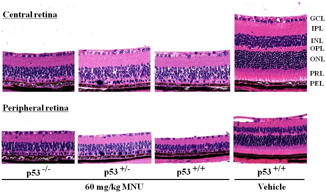
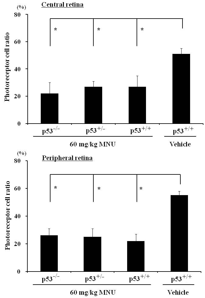
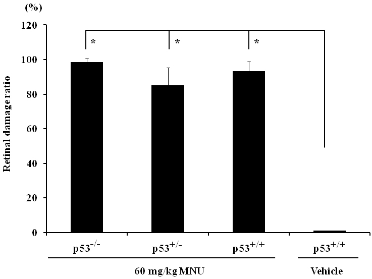
Methods: Eight-week-old p53(-/-), p53(+/-), and p53(+/+) mice received an intraperitoneal injection of 60 mg/kg bodyweight of MNU. Age-matched p53(+/+) mice received the vehicle only (physiologic saline containing 0.05% acetic acid). Mice were sacrificed and necropsied 7 days after the treatment. Both eyes were examined histologically and morphometrically to determine retinal thickness, photoreceptor cell ratio, and retinal damage ratio.
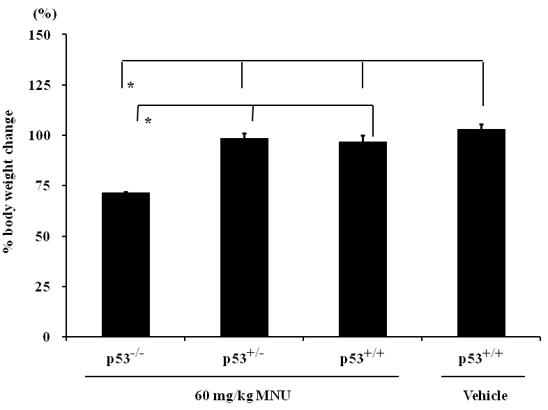
Results: No mice died during the experiment, but the p53 null mice treated with MNU had a statistically significant weight loss compared to the other groups. Histologically, all MNU-treated mice, regardless of p53 gene status, experienced retinal degeneration characterized by photoreceptor cell loss (the disappearance of the outer nuclear layer and photoreceptor layer) in both the central and peripheral retina. All MNU-treated mice had significantly decreased retinal thickness and photoreceptor cell ratios at the central and peripheral retina and an increased retinal damage ratio compared to the vehicle-treated control. The retinal changes caused by MNU in p53(+/+), p53(+/-), and p53(-/-) mice were not significantly different and hence were related to a p53-independent apoptotic mechanism.
Conclusions: Because the absence of p53 did not prevent photoreceptor cell loss, we conclude that p53 is not essential for MNU-mediated photoreceptor cell degeneration.
Figures
References
-
- Hartong DT, Berson EL, Dryja TP. Retinitis pigmentosa. Lancet. 2006;368:1795–809. - PubMed
-
- Shintani K, Shechtman DL, Gurwood AS. Review and update: current treatment trends for patients with retinitis pigmentosa. Optometry. 2009;80:384–401. - PubMed
-
- Sancho-Pelluz J, Arango-Gonzalez B, Kustermann S, Romero FJ, van Veen T, Zrenner E, Ekström P, Paquet-Durand F. Photoreceptor cell death mechanisms in inherited retinal degeneration. Mol Neurobiol. 2008;38:253–69. - PubMed
-
- Rivas MA, Vecino E. Animal models and different therapies for treatment of retinitis pigmentosa. Histol Histopathol. 2009;24:1295–322. - PubMed
-
- Tsubura A, Yuri T, Yoshizawa K, Uehara N, Takada H. Role of fatty acids in malignancy and visual impairment: epidemiological evidence and experimental studies. Histol Histopathol. 2009;24:223–34. - PubMed
Publication types
MeSH terms
Substances
LinkOut - more resources
Full Text Sources
Molecular Biology Databases
Research Materials
Miscellaneous