

Towards the sensory nature of the carotid body: hering, de castro and heymansdagger
- PMID: 20057927
- PMCID: PMC2802533
- DOI: 10.3389/neuro.05.023.2009
Towards the sensory nature of the carotid body: hering, de castro and heymansdagger
Abstract
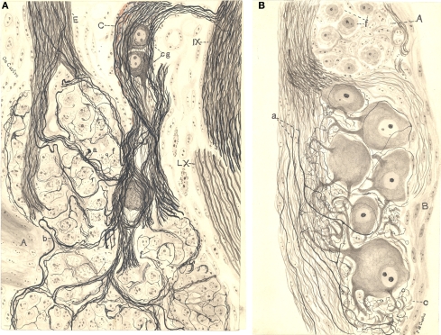
The carotid body or glomus caroticum is a chemosensory organ bilaterally located between the external and internal carotid arteries. Although known by anatomists since the report included by Von Haller and Taube in the mid XVIII century, its detailed study started the first quarter of the XX. The Austro-German physiologist Heinrich E. Hering studied the cardio-respiratory reflexes searched for the anatomical basis of this reflex in the carotid sinus, while the Ghent School leaded by the physio-pharmacologists Jean-François Heymans and his son Corneille focussed in the cardio-aortic reflexogenic region. In 1925, Fernando De Castro, one of the youngest and more brilliant disciples of Santiago Ramón y Cajal at the Laboratorio de Investigaciones Biológicas (Madrid, Spain), profited from some original novelties in histological procedures to study the fine structure and innervation of the carotid body. De Castro unravelled them in a series of scientific papers published between 1926 and 1929, which became the basis to consider the carotid body as a sensory receptor (or chemoreceptor) to detect the chemical changes in the composition of the blood. Indeed, this was the first description of arterial chemoreceptors. Impressed by the novelty and implications of the work of De Castro, Corneille Heymans invited the Spanish neurologist to visit Ghent on two occasions (1929 and 1932), where both performed experiences together. Shortly after, Heymans visited De Castro at the Instituto Cajal (Madrid). From 1932 to 1933, Corneille Heymans focused all his attention on the carotid body his physiological demonstration of De Castro's hypothesis regarding chemoreceptors was awarded with the Nobel Prize in Physiology or Medicine in 1938, just when Spain was immersed in its catastrophic Civil War.
Keywords: baroreceptor; blood circulation; carotid body; chemoreceptor; history of neuroscience; nobel prize; physiology; respiratory reflex.
Figures


Similar articles
-
The discovery of sensory nature of the carotid bodies--invited article.Adv Exp Med Biol. 2009;648:1-18. doi: 10.1007/978-90-481-2259-2_1. Adv Exp Med Biol. 2009. PMID: 19536460
-
Fernando de Castro: Cajal's Man on the Peripheral Nervous System.Anat Rec (Hoboken). 2020 May;303(5):1206-1214. doi: 10.1002/ar.24191. Epub 2019 Jun 20. Anat Rec (Hoboken). 2020. PMID: 31172650
-
Fernando de Castro and the discovery of the arterial chemoreceptors.Front Neuroanat. 2014 May 12;8:25. doi: 10.3389/fnana.2014.00025. eCollection 2014. Front Neuroanat. 2014. PMID: 24860435 Free PMC article. Review.
-
The Cajal School in the Peripheral Nervous System: The Transcendent Contributions of Fernando de Castro on the Microscopic Structure of Sensory and Autonomic Motor Ganglia.Front Neuroanat. 2016 Apr 20;10:43. doi: 10.3389/fnana.2016.00043. eCollection 2016. Front Neuroanat. 2016. PMID: 27147984 Free PMC article. Review.
-
Cajal and the Spanish Neurological School: Neuroscience Would Have Been a Different Story Without Them.Front Cell Neurosci. 2019 May 24;13:187. doi: 10.3389/fncel.2019.00187. eCollection 2019. Front Cell Neurosci. 2019. PMID: 31178695 Free PMC article. Review.
Cited by
-
The Women Neuroscientists in the Cajal School.Front Neuroanat. 2019 Jul 16;13:72. doi: 10.3389/fnana.2019.00072. eCollection 2019. Front Neuroanat. 2019. PMID: 31379519 Free PMC article.
-
Tentonin 3 as a baroreceptor mechanosensor: not a stretch.J Clin Invest. 2020 Jul 1;130(7):3412-3415. doi: 10.1172/JCI138120. J Clin Invest. 2020. PMID: 32484454 Free PMC article.
-
Peripheral chemoreceptors: function and plasticity of the carotid body.Compr Physiol. 2012 Jan;2(1):141-219. doi: 10.1002/cphy.c100069. Compr Physiol. 2012. PMID: 23728973 Free PMC article. Review.
-
The carotid body: A novel key player in neuroimmune interactions.Front Immunol. 2022 Oct 24;13:1033774. doi: 10.3389/fimmu.2022.1033774. eCollection 2022. Front Immunol. 2022. PMID: 36389846 Free PMC article. Review.
-
Carotid body chemoreceptors: physiology, pathology, and implications for health and disease.Physiol Rev. 2021 Jul 1;101(3):1177-1235. doi: 10.1152/physrev.00039.2019. Epub 2021 Feb 11. Physiol Rev. 2021. PMID: 33570461 Free PMC article. Review.
References
-
- Comroe J. H. (1939). The location and function of the chemoreceptors of the aorta. Am. J. Physiol. 127, 176–191
-
- Cyon E., Ludwig C. (1866). Die Reflexe eines der sensiblen Nerven des Herzens auf die Motorischen der Blutgefässe. Ber. Sächs. Ges. Wiss. Leipzig. 18, 307–329
-
- De Castro F. (1921). Estudio sobre los ganglios sensitivos del hombre adulto en estado normal y patológico. Trab. Lab. Invest. Biol. 19, 241–340
-
- De Castro F. (1922). Evolución de los ganglios simpáticos vertebrales y prevertebrales. Conexiones y citoarquitectonia en algunos grupos de ganglio en el niño y en el hombre adulto. Trab. Lab. Invest. Biol. 20, 113–208
-
- De Castro F. (1925). Technique pour la coloration du système nerveux quand il est pourvu de ses étuis osseux. Trav. Lab. Rech. Biol. 23, 427–446
LinkOut - more resources
Full Text Sources