. 2009 Nov 20;10(10):4352-4374.
doi: 10.3390/ijms10104352.

Improved adhesion, growth and maturation of vascular smooth muscle cells on polyethylene grafted with bioactive molecules and carbon particles

Affiliations

Improved adhesion, growth and maturation of vascular smooth muscle cells on polyethylene grafted with bioactive molecules and carbon particles

Martin Parizek et al. Int J Mol Sci. .

Abstract

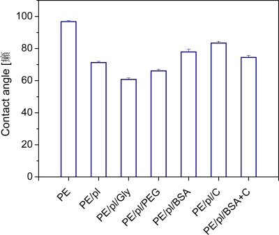
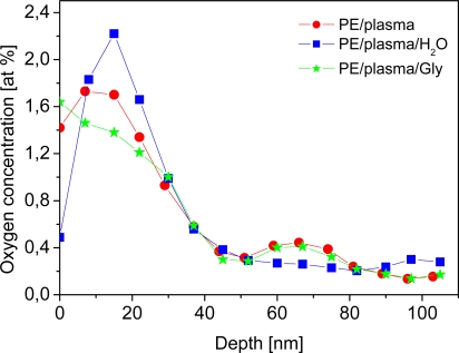
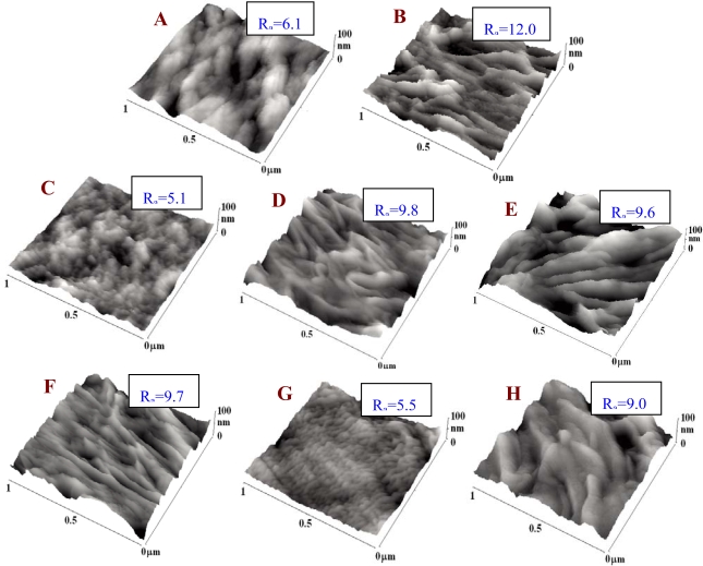
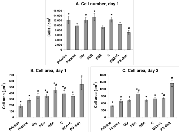
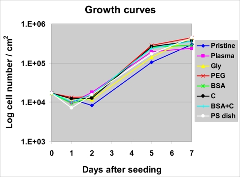
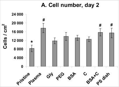
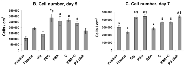
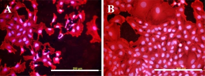
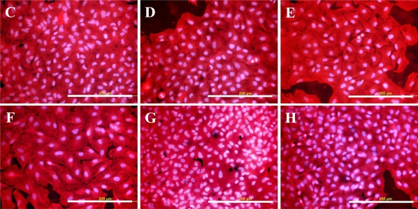
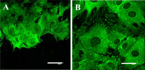
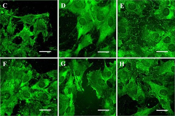
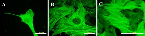
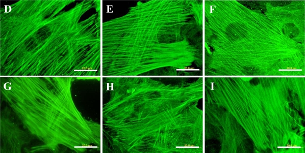
High-density polyethylene (PE) foils were modified by an Ar(+) plasma discharge and subsequent grafting with biomolecules, namely glycine (Gly), polyethylene glycol (PEG), bovine serum albumin (BSA), colloidal carbon particles (C) or BSA and C (BSA + C). As revealed by atomic force microscopy (AFM), goniometry and Rutherford Backscattering Spectroscopy (RBS), the surface chemical structure and surface morphology of PE changed dramatically after plasma treatment. The contact angle decreased for the samples treated by plasma, mainly in relation to the formation of oxygen structures during plasma irradiation. A further decrease in the contact angle was obvious after glycine and PEG grafting. The increase in oxygen concentration after glycine and PEG grafting proved that the two molecules were chemically linked to the plasma-activated surface. Plasma treatment led to ablation of the PE surface layer, thus the surface morphology was changed and the surface roughness was increased. The materials were then seeded with vascular smooth muscle cells (VSMC) derived from rat aorta and incubated in a DMEM medium with fetal bovine serum. Generally, the cells adhered and grew better on modified rather than on unmodified PE samples. Immunofluorescence showed that focal adhesion plaques containing talin, vinculin and paxillin were most apparent in cells on PE grafted with PEG or BSA + C, and the fibres containing alpha-actin, beta-actin or SM1 and SM2 myosins were thicker, more numerous and more brightly stained in the cells on all modified PE samples than on pristine PE. An enzyme-linked immunosorbent assay (ELISA) revealed increased concentrations of focal adhesion proteins talin and vinculin and also a cytoskeletal protein beta-actin in cells on PE modified with BSA + C. A contractile protein alpha-actin was increased in cells on PE grafted with PEG or Gly. These results showed that PE activated with plasma and subsequently grafted with bioactive molecules and colloidal C particles, especially with PEG and BSA + C, promotes the adhesion, proliferation and phenotypic maturation of VSMC.

Keywords: bioactivity; biocompatibility; plasma irradiation; tissue engineering and reconstruction.


Figures

Figure 1.
Figure 1.
The contact angle of pristine PE (PE), plasma-treated PE (PE/pl), PE plasma-treated and then grafted with glycine (PE/pl/Gly), PEG (PE/pl/PEG), BSA (PE/pl/BSA), C (PE/pl/C) or BSA + C (PE/pl/BSA + C).
Figure 2.
Figure 2.
Depth concentration profile of oxygen, determined from RBS measurement, on the sample exposed to plasma discharge (PE/plasma), plasma-treated sample immersed in water (PE/plasma/H2O) and plasma-treated sample grafted with glycine (PE/plasma/Gly).
Figure 3.
Figure 3.
AFM images of pristine PE (A), plasma-treated PE (B), PE treated with plasma and immersed in water (C), PE treated with plasma and grafted with glycine (D), PEG (E), BSA (F), colloidal carbon particles (G) or BSA + C (H). Ra is a parameter of measured surface roughness in nm.
Figure 4.
Figure 4.
The number of initially adhered cells (A) and the size of the cell spreading area (B, C) of rat aortic smooth muscle cells in cultures on non-modified PE (Pristine), PE irradiated with plasma (Plasma), PE irradiated with plasma and grafted with glycine (Gly), polyethyleneglycol (PEG) bovine serum albumin (BSA), colloidal carbon particles (C) or bovine serum albumin and C (BSA + C). As a reference material, a tissue culture polystyrene dish (PS dish) was used. Means ± S.E.M. from 3 samples, each measured for 50 times (A) or from 72 to 142 cells for each experimental group (C, D). ANOVA, Student-Newman-Keuls method. Statistical significance: *, #: p ≤ 0.05 compared to the value on pristine PE and polystyrene dish, respectively.
Figure 5.
Figure 5.
Growth dynamics of rat aortic smooth muscle cells in cultures on non-modified PE (Pristine), PE irradiated with plasma (Plasma), PE irradiated with plasma and grafted with glycine (Gly), polyethyleneglycol (PEG) bovine serum albumin (BSA), colloidal carbon particles (C) or bovine serum albumin and C (BSA + C). As a reference material, a tissue culture polystyrene dish (PS dish) was used. Means from three samples for each experimental group and time interval (each sample measured for 50 times).
Figure 6.
Figure 6.
Proliferation activity of rat aortic smooth muscle cells measured by cell numbers achieved on day 2 (A), 5 (B) and 7 (C) after seeding on non-modified PE (Pristine), PE irradiated with plasma (Plasma), PE irradiated with plasma and grafted with glycine (Gly), polyethyleneglycol (PEG) bovine serum albumin (BSA), colloidal carbon particles (C) or bovine serum albumin and C (BSA + C). As a reference material, a tissue culture polystyrene dish (PS dish) was used. Means ± S.E.M. from three samples for each experimental group, each measured for 50 times. ANOVA, Student-Newman-Keuls method. Statistical significance: *,#, $: p ≤ 0.05 compared to the value on pristine PE, polystyrene dish and PE irradiated with plasma, respectively.
Figure 6.
Figure 6.
Proliferation activity of rat aortic smooth muscle cells measured by cell numbers achieved on day 2 (A), 5 (B) and 7 (C) after seeding on non-modified PE (Pristine), PE irradiated with plasma (Plasma), PE irradiated with plasma and grafted with glycine (Gly), polyethyleneglycol (PEG) bovine serum albumin (BSA), colloidal carbon particles (C) or bovine serum albumin and C (BSA + C). As a reference material, a tissue culture polystyrene dish (PS dish) was used. Means ± S.E.M. from three samples for each experimental group, each measured for 50 times. ANOVA, Student-Newman-Keuls method. Statistical significance: *,#, $: p ≤ 0.05 compared to the value on pristine PE, polystyrene dish and PE irradiated with plasma, respectively.
Figure 7.
Figure 7.
Morphology of rat aortic smooth muscle cells on day 5 after seeding on pristine PE (A), tissue culture polystyrene dish (B), PE irradiated with Ar+ plasma (C), PE irradiated with plasma and grafted with glycine (D), polyethyleneglycol (E) bovine serum albumin (F), colloidal carbon particles (G) or bovine serum albumin and C (H). Cell membrane and cytoplasm stained with Texas Red C2-maleimide (red fluorescence), cell nuclei with Hoechst #33342 (blue fluorescence). Olympus IX 51 microscope, DP 70 digital camera, obj. 20×, bar = 200 μm.
Figure 7.
Figure 7.
Morphology of rat aortic smooth muscle cells on day 5 after seeding on pristine PE (A), tissue culture polystyrene dish (B), PE irradiated with Ar+ plasma (C), PE irradiated with plasma and grafted with glycine (D), polyethyleneglycol (E) bovine serum albumin (F), colloidal carbon particles (G) or bovine serum albumin and C (H). Cell membrane and cytoplasm stained with Texas Red C2-maleimide (red fluorescence), cell nuclei with Hoechst #33342 (blue fluorescence). Olympus IX 51 microscope, DP 70 digital camera, obj. 20×, bar = 200 μm.
Figure 8.
Figure 8.
Immunofluorescence staining of talin, an integrin-associated protein of focal adhesion plaques, in rat aortic smooth muscle cells on day 5 after seeding on pristine PE (A), a reference material represented by a microscopic glass coverslip (B), PE irradiated with plasma (C), PE irradiated with plasma and grafted with glycine (D), polyethyleneglycol (E) bovine serum albumin (F), colloidal carbon particles (G) or bovine serum albumin and C (H). Leica confocal laser scanning microscope (TCS SP2, Germany), obj. 100x, bar = 30 μm.
Figure 8.
Figure 8.
Immunofluorescence staining of talin, an integrin-associated protein of focal adhesion plaques, in rat aortic smooth muscle cells on day 5 after seeding on pristine PE (A), a reference material represented by a microscopic glass coverslip (B), PE irradiated with plasma (C), PE irradiated with plasma and grafted with glycine (D), polyethyleneglycol (E) bovine serum albumin (F), colloidal carbon particles (G) or bovine serum albumin and C (H). Leica confocal laser scanning microscope (TCS SP2, Germany), obj. 100x, bar = 30 μm.
Figure 9.
Figure 9.
Immunofluorescence staining of contractile protein alpha-actin in rat aortic smooth muscle cells on day 5 after seeding on pristine PE (A), reference materials represented by a microscopic glass coverslip (B), a standard cell culture polystyrene dish (C), PE irradiated with plasma (D), PE irradiated with plasma and grafted with glycine (E), polyethyleneglycol (F), bovine serum albumin (G), colloidal carbon particles (H) or bovine serum albumin and C (I). Olympus IX 51 microscope, DP 70 digital camera, obj. 100× (A, B, D-I) or 40× (C). Bar = 20 μm (A, B, D-I) or 100 μm (C).
Figure 9.
Figure 9.
Immunofluorescence staining of contractile protein alpha-actin in rat aortic smooth muscle cells on day 5 after seeding on pristine PE (A), reference materials represented by a microscopic glass coverslip (B), a standard cell culture polystyrene dish (C), PE irradiated with plasma (D), PE irradiated with plasma and grafted with glycine (E), polyethyleneglycol (F), bovine serum albumin (G), colloidal carbon particles (H) or bovine serum albumin and C (I). Olympus IX 51 microscope, DP 70 digital camera, obj. 100× (A, B, D-I) or 40× (C). Bar = 20 μm (A, B, D-I) or 100 μm (C).

Similar articles

Cited by

References

    1. Bacakova L, Svorcik V, Rybka V, Micek I, Hnatowicz V, Lisa V, Kocourek F. Adhesion and proliferation of cultured human vascular smooth muscle cells on polystyrene implanted with N+, F+ and Ar+ ions. Biomaterials. 1996;17:1121–1126. - PubMed
    1. Bacakova L, Mares V, Bottone MG, Pellicciari C, Lisa V, Svorcik V. Fluorine-ion-implanted polystyrene improves growth and viability of vascular smooth muscle cells in culture. J. Biomed. Mater. Res. 2000;49:369–379. - PubMed
    1. Bacakova L, Walachova K, Svorcik V, Hnatowicz V. Adhesion and proliferation of rat vascular smooth muscle cells on polyethylene implanted with O+ and C+ ions. J. Biomater. Sci.—Polym. Ed. 2001;12:817–834. - PubMed
    1. Bacakova L, Filova E, Rypacek F, Svorcik V, Stary V. Cell adhesion on artificial materials for tissue engineering. Physiol. Res. 2004;53:S35–S45. - PubMed
    1. Bacakova L, Svorcik V. Cell colonization control by physical and chemical modification of materials. In: Kimura D, editor. Cell Growth Processes: New Research. Nova Science Publishers, Inc.; Hauppauge, NY, USA: 2008. pp. 5–56.

Publication types

MeSH terms