

Review
doi: 10.1007/s10554-009-9571-x.
Quantification in cardiac MRI: advances in image acquisition and processing
Affiliations
- PMID: 20058082
- PMCID: PMC2816803
- DOI: 10.1007/s10554-009-9571-x
Item in Clipboard
Review
Quantification in cardiac MRI: advances in image acquisition and processing
Int J Cardiovasc Imaging.
2010 Feb.
Abstract
Cardiac magnetic resonance (CMR) imaging enables accurate and reproducible quantification of measurements of global and regional ventricular function, blood flow, perfusion at rest and stress as well as myocardial injury. Recent advances in MR hardware and software have resulted in significant improvements in image quality and a reduction in imaging time. Methods for automated and robust assessment of the parameters of cardiac function,blood flow and morphology are being developed. This article reviews the recent advances in image acquisition and quantitative image analysis in CMR.
Figures
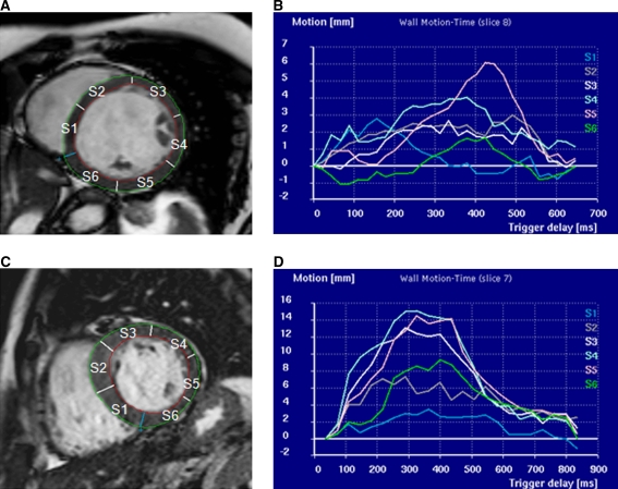
a, b Patient with dyssynchronous myocardial wall motion as clearly revealed by the uncoordinated wall motion pattern derived from the endocardial contours detected in every image frame. c, d Patient with normal synchronous wall motion as indicated by the normal temporal wall motion curve
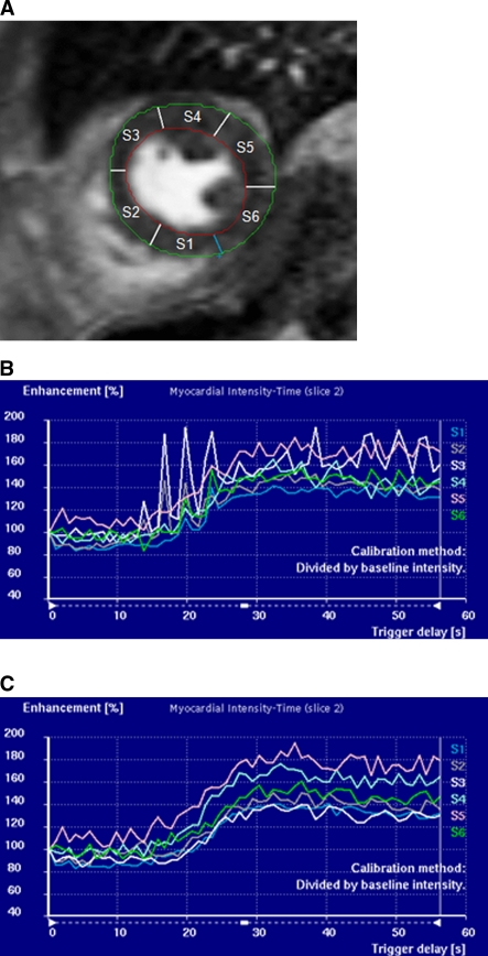
Short-axis image at a mid ventricular slice level with endo- and epi-cardial contours defined and the myocardium divided into 6 segments (a). Signal-intensity versus time curves are derived for each of the myocardial segments. Without motion correction (b) the curves are not suitable for quantitative analysis. After motion correction (c), perfusion indices such as maximum upslope can be derived reliably
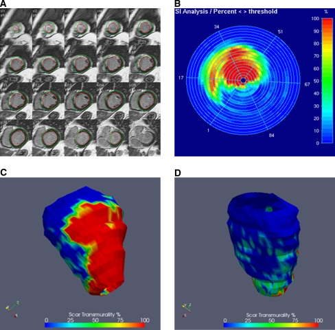
a Multi-slice short-axis DEMR with defined endocardial and epicardial contours superimposed. Scar transmurality defined as the thickness of scar relative to the local wall thickness assessed from DEMR. Scar distribution can be displayed as a bulls-eye plot (b), or as three-dimensional reconstructions (c, d)
References
Publication types
MeSH terms
LinkOut - more resources
Full Text Sources
Other Literature Sources
Medical
