Review
. 2010 Jan;91(1):1-19.
doi: 10.1007/s12185-009-0476-1.

Pathophysiology of thrombotic thrombocytopenic purpura

Affiliations
Review

Pathophysiology of thrombotic thrombocytopenic purpura

Han-Mou Tsai. Int J Hematol. 2010 Jan.

Abstract

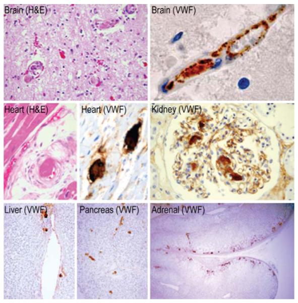
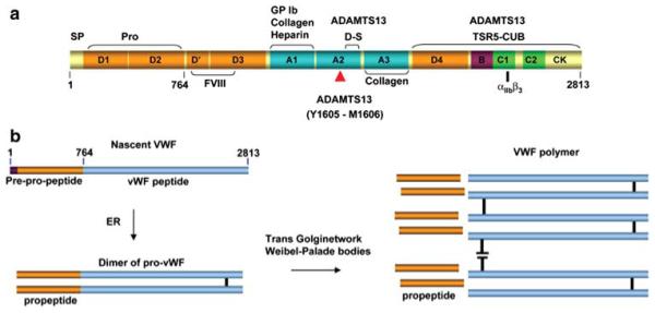
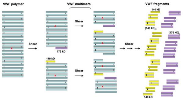
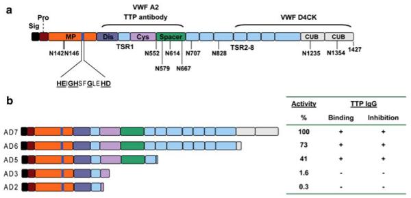
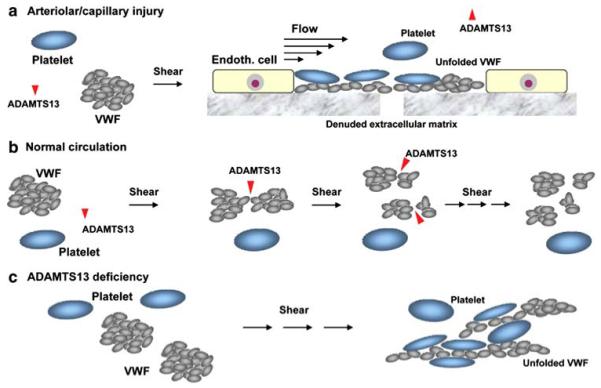
Thrombotic thrombocytopenic purpura (TTP) is a disorder with characteristic von Willebrand factor (VWF)-rich microthrombi affecting the arterioles and capillaries of multiple organs. The disorder frequently leads to early death unless the patients are treated with plasma exchange or infusion. Studies in the last decade have provided ample evidence to support that TTP is caused by deficiency of a plasma metalloprotease, ADAMTS13. When exposed to high shear stress in the microcirculation, VWF and platelets are prone to form aggregates. This propensity of VWF and platelet to form microvascular thrombosis is mitigated by ADAMTS13, which cleaves VWF before it is activated by shear stress to cause platelet aggregation in the circulation. Deficiency of ADAMTS13, due to autoimmune inhibitors in patients with acquired TTP and mutations of the ADAMTS13 gene in hereditary cases, leads to VWF-platelet aggregation and microvascular thrombosis of TTP. In this review, we discuss the current knowledge on the pathogenesis, diagnosis and management of TTP, address the ongoing controversies, and indicate the directions of future investigations.


Figures

Fig. 1
Fig. 1
Immunohistopathology of TTP. VWF-rich hyaline thrombi are present in the arterioles and capillaries but not the venules of multiple organs of a patient that died of TTP due to ADAMTS13 inhibitors. The endothelial cells are intact and contain abundant granules of VWF. No inflammatory cell infiltration is noted. The renal glomeruli contain foci of microthrombi but have well preserved architectures, consistent with the patient’s hematuria and minimal Cr elevation (1.2 mg/dL) at the time of death. VWF is not expressed in the sinusoidal endothelial cells of the liver and no thrombi are detected in the hepatic sinusoids
Fig. 2
Fig. 2
The biosynthesis and the domain structure of VWF. a The domain structure of VWF. The location of the Y1605-M1606 scissile bond and the regions of the A2 domain and the D4-CK domains that are involved in binding with ADAMTS13 exocites (distintegrin-spacer domains [D-S] and TSR5-CUB domains, respectively) are indicated. Also shown are the regions of the VWF polypeptide that are involved in binding factor VIII, GP1b, collagen, heparin and GPαIIbβ3. b Formation of VWF polymers. In the endoplasmic reticulum (ER), two pro-VWF polypeptides are linked by disulfide bonds near the C terminus to form a dimer of pro-VWF. The dimers are bonded by disulfide bonds near the N terminus to form a polymer for secretion from endothelial cells. The process of polymerization starts in the trans Golgi network and proceeds to completion in the Weibel-Palade bodies. The propeptide is cleaved before the VWF polymer is secreted
Fig. 3
Fig. 3
Generation of VWF multimers. At each brief exposure to high shear stress during the transit through the arteriole/capillary microcirculation, a VWF polymer or multimer is expected to be cleaved by ADAMTS13 at one of its Y1605-M1606 bonds at or near its center subunit (red, filled triangle). A series of multimers with progressively smaller sizes are generated when this process is repeated on the cleaved products. The smallest products of this proteolysis are homodimers of the 140 and the 176 kD fragments. The 140 kD monomer has not yet been detected in normal plasma. Only the VWF fragments would be generated if a VWF polymer were fully unfolded and cleaved simultaneously at all the scissile bonds
Fig. 4
Fig. 4
The structure and function of ADAMTS13. a ADAMTS13 has a modular structure comprising of a signal peptide (Sig), a propeptide (Pro), a metalloprotease domain (MP), a disintergrin-like domain (Dis), a thrombospondin type 1 repeat (TSR), a cysteine-rich region (Cys), a spacer domain (Spa), 7 additional TSRs (TSR 2-8), and two CUB domains. The 10 glycosylated N residues are indicated. The catalytic site (HEIGHSFGLEHD) is in the metalloprotease domain. The underline residues are conserved in the ADAMTS metalloprotease family. The sequence between the disintegrin and spacer domains interacts with regions of the VWF A2 domain downstream of the scissile bond. The sequence between TSR5 and CUB domains interacts with an epitope in the D4-CK region of VWF. This latter interaction has modest affinity and does not require shear stress. However, it may promote the other exocite interaction and subsequent Y1605-M1606 cleavage. b A comparison of ADAMTS13 with its 4 truncated variants for their VWF cleaving activity and binding and suppression by the IgG inhibitors of TTP patients
Fig. 5
Fig. 5
ADAMTS13 prevents VWF–platelet aggregation and thrombosis. a At sites of vessel injury under high shear stress conditions such as those in the arterioles and capillaries, the conformationally flexible VWF, upon attached to the extracellular matrix of the denuded subendothelium, is readily stretched to an extended form for mediating platelet adhesion and aggregation essential for hemostasis. The unfolded VWF may be protected by thrombospondin-1 from ADAMTS13 cleavage. Inactivation of ADAMTS13 by thrombin and plasmin generated at sites of vessel injury may also help minimize the cleavage of the unfolded VWF. b In the circulation, the stretching of VWF does not occur because ADAMTS13 cleaves the Y1605-M1606 bond of the A2 domain whenever it is exposed by shear stress. The cleavage occurs each time VWF traverses the microcirculation. The proposed scheme assumes that VWF is cleaved when its structural changes occur at the domain level, before it becomes stretched by shear stress to an elongated form. This process of proteolysis maintains the VWF in a compact form while it is converted to a series of multimers with progressively smaller sizes. c In the absence of ADAMTS13, VWF, after repetitive exposure to high levels of shear stress, is unfolded to an elongated active form that causes platelet aggregation in the circulation. Intra-vascular platelet thrombosis further increases the shear stress in the microcirculation, begetting more VWF unfolding and platelet aggregation. This self-perpetuating process may explain why TTP often deteriorates precipitously if it is not immediately treated with plasma therapy to replenish the missing ADAMTS13

Similar articles

Cited by

References

    1. Asada Y, Sumiyoshi A, Hayashi T, Suzumiya J, Kaketani K. Immunohistochemistry of vascular lesion in thrombotic thrombocytopenic purpura, with special reference to factor VIII related antigen. Thromb Res. 1985;38:469–79. - PubMed
    1. Tsai HM, Chandler WL, Sarode R, et al. von Willebrand factor and von Willebrand factor-cleaving metalloprotease activity in Escherichia coli O157:H7-associated hemolytic uremic syndrome. Pediatr Res. 2001;49:653–9. - PubMed
    1. Hosler GA, Cusumano AM, Hutchins GM. Thrombotic thrombocytopenic purpura and hemolytic uremic syndrome are distinct pathologic entities. A review of 56 autopsy cases. Arch Pathol Lab Med. 2003;127:834–9. - PubMed
    1. Sadler JE. New concepts in von Willebrand disease. Annu Rev Med. 2005;56:173–91. - PubMed
    1. Tsai HM, Nagel RL, Hatcher VB, Sussman II. Multimeric composition of endothelial cell-derived von Willebrand factor. Blood. 1989;73:2074–6. - PubMed

Publication types

MeSH terms

LinkOut - more resources