. 2010 Jun;28(6):703-9.
doi: 10.1002/jor.21071.

Collagen scaffold supplementation does not improve the functional properties of the repaired anterior cruciate ligament

Affiliations

Collagen scaffold supplementation does not improve the functional properties of the repaired anterior cruciate ligament

Braden C Fleming et al. J Orthop Res. 2010 Jun.

Abstract

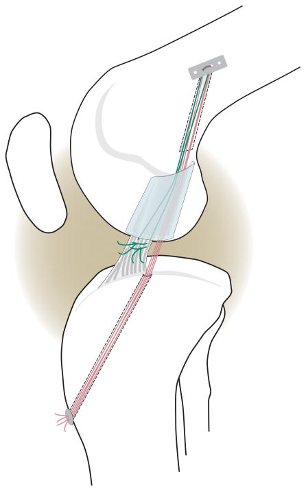
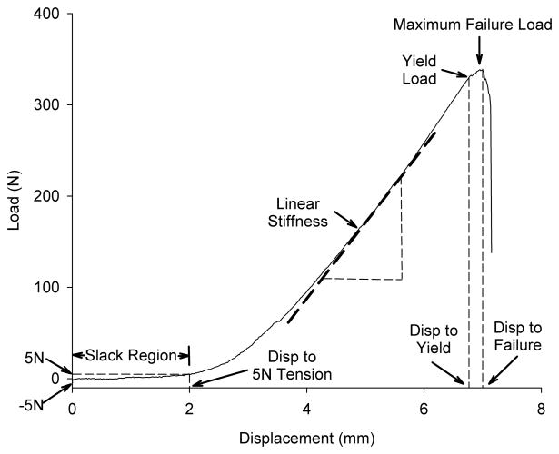
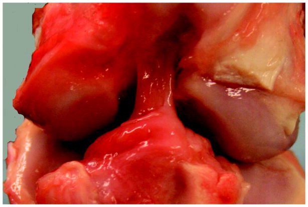
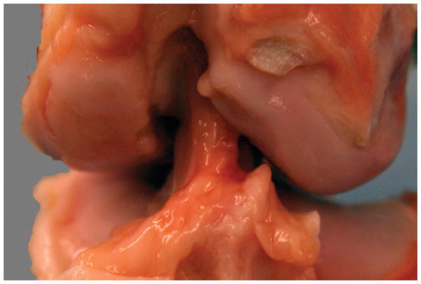
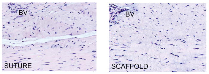
Primary suture anterior cruciate ligament (ACL) repair was abandoned in favor of reconstruction due to a high rate of clinical failures. However, the insertion of a collagen scaffold loaded with platelets into the wound at the time of suture repair ("enhanced primary repair") has been shown to improve functional healing in animal models. Our objectives were to determine if using a collagen scaffold alone (without platelets) would be sufficient to increase the structural properties of the repaired ACL and decrease postoperative knee laxity compared to suture repair without the scaffold. Eight Yucatan minipigs underwent bilateral ACL transection and suture repair. In one knee, the repair was augmented with a collagen scaffold (SCAFFOLD group) while the other had suture alone (SUTURE group). After 13 weeks of healing, knee joint laxity and the structural properties of the ACL were measured. The addition of the collagen scaffold to suture repair of a transected ACL did not significantly improve the mean anteroposterior knee laxity [SCAFFOLD vs. SUTURE: 6.1 +/- 1.4 vs. 4.4 +/- 2.0 mm (p = 0.07), 8.1 +/- 2.0 vs. 7.6 +/- 2.0 mm (p = 0.66), and 6.2 +/- 1.2 vs. 6.1 +/- 1.8 mm (p = 0.85) at 30 degrees, 60 degrees, and 90 degrees flexion, respectively]. Likewise, there were no significant differences in the structural properties [SCAFFOLD vs. SUTURE: 367 +/- 185.9 vs. 322 +/- 122.0N (p = 0.66) and 90.7 +/- 29.5 vs. 85.0 +/- 30.3N/mm (p = 0.74) for the yield load and linear stiffness, respectively]. The use of a collagen scaffold alone to enhance suture repair of the ACL was ineffective in this animal model. Future work will be directed at stimulating biological activity in the scaffold.


Figures

Fig. 1
Fig. 1
Schematic diagram depicting the primary suture repair with the collagen scaffold in place. Sutures were fixed proximally with an Endobutton. The sponge was threaded onto four of the trailing suture ends (RED) which were then passed through the tibial tunnel and tied over a button to provide initial knee stability. The remaining two suture ends (GREEN) were tied to the sutures in the tibial stump of the ACL. All sutures were resorbable, and there was no sign of suture material remaining at the time of post-mortem testing.
Fig. 2
Fig. 2
Schematic of the load-displacement curve for tensile testing of the femur-ACL-tibia complex. The toe region (the displacement of the joint surfaces at low loads) was defined by the displacement between −5 and +5N of tensile load. After the slack of the ligament was removed, the slope increased as it entered the linear stiffness region until the yield load was reached. Maximum failure load was defined as the maximum load supported by the construct. Yield load is defined as the location on the load-displacement curve where the slope of the curve first deviates by 2% from the linear region. The displacements to yield and failure were defined as the amount of excursion of joint surfaces at the yield load and the maximum failure load, respectively.
Figure 3
Figure 3
Figure 3A: Gross appearance of a knee treated with SUTURE repair alone. Note the repair tissue arising from just posterior to the fat pad, from the anterior slope of the tibial spines, and coursing from the tibia to the lateral wall of the intercondylar notch. There is no significant change noted in the femoral condylar cartilage either medially or laterally. The repair tissue itself is composed of multiple individual synovialized bands. Figure 3B. Gross appearance of a knee treated with SCAFFOLD supplementation of the suture repair. Note the similar appearance with the SUTURE knee in 3A. Again, there is repair tissue coursing from tibia to femur in one continuous mass. There is no change noted in the femoral condylar cartilage. The repair tissue itself is composed of multiple individual synovialized bands.
Figure 3
Figure 3
Figure 3A: Gross appearance of a knee treated with SUTURE repair alone. Note the repair tissue arising from just posterior to the fat pad, from the anterior slope of the tibial spines, and coursing from the tibia to the lateral wall of the intercondylar notch. There is no significant change noted in the femoral condylar cartilage either medially or laterally. The repair tissue itself is composed of multiple individual synovialized bands. Figure 3B. Gross appearance of a knee treated with SCAFFOLD supplementation of the suture repair. Note the similar appearance with the SUTURE knee in 3A. Again, there is repair tissue coursing from tibia to femur in one continuous mass. There is no change noted in the femoral condylar cartilage. The repair tissue itself is composed of multiple individual synovialized bands.
Figure 4
Figure 4
Representative histomicrographs of healing ligament from the SUTURE and SPONGE groups. Note that the healing ligaments had areas of parallel collagen fascicles (bottom of both micrographs) as well as more irregular areas (top of both micrographs). BV= Blood vessel. Hematoxylin and Eosin, 200× magnification.

Similar articles

Cited by

References

    1. Praemer A, Furner S, Rice DP. Musculoskeletal Conditions in the United States. American Academy of Orthopaedic Surgeons 1999
    1. Adriani E, Mariani PP, Maresca G, Santori N. Healing of the patellar tendon after harvesting of its mid-third for anterior cruciate ligament reconstruction and evolution of the unclosed donor site defect. Knee Surg Sports Traumatol Arthrosc. 1995;3:138–143. - PubMed
    1. Cabaud HE, Rodkey WG, Feagin JA. Experimental studies of acute anterior cruciate ligament injury and repair. Am J Sports Med. 1979;7:18–22. - PubMed
    1. O’Donoghue DH, Rockwood C, Frank GR, Jack SC, Kenyon R. Repair of the anterior cruciate ligament in dogs. J Bone Joint Surg [Am] 1966;48:503–519. - PubMed
    1. O’Donoghue DH, Frank GR, Jeter GL, Johnson W, Zeiders JW, Kenyon R. Repair and reconstruction of the anterior cruciate ligament in dogs. Factors influencing long-term results. J Bone Joint Surg [Am] 1971;53:710–718. - PubMed

Publication types