. 2010 Mar 15;518(6):800-14.
doi: 10.1002/cne.22243.

A novel model of retinal ablation demonstrates that the extent of rod cell death regulates the origin of the regenerated zebrafish rod photoreceptors

Affiliations

A novel model of retinal ablation demonstrates that the extent of rod cell death regulates the origin of the regenerated zebrafish rod photoreceptors

Jacob E Montgomery et al. J Comp Neurol. .

Abstract

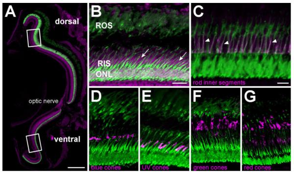
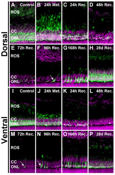
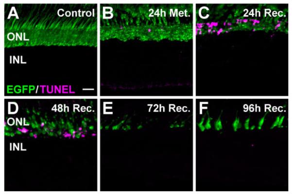
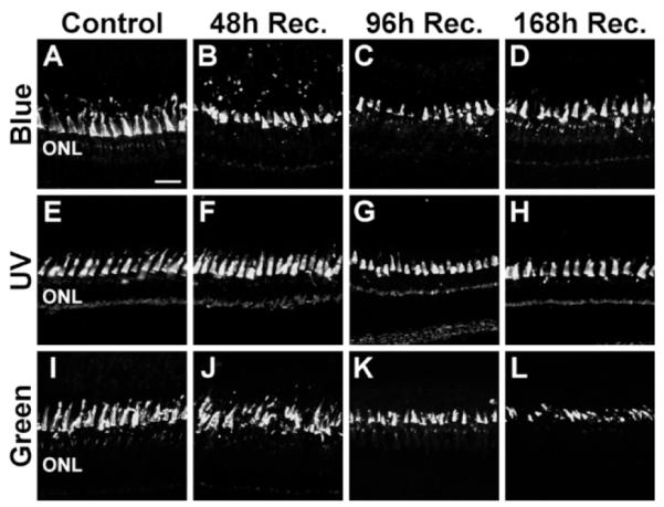
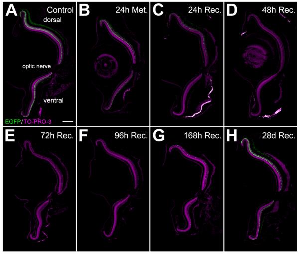
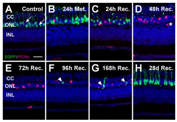
The adult zebrafish retina continuously produces rod photoreceptors from infrequent Müller glial cell division, yielding neuronal progenitor cells that migrate to the outer nuclear layer and become rod precursor cells that are committed to differentiate into rods. Retinal damage models suggested that rod cell death induces regeneration from rod precursor cells, whereas loss of any other retinal neurons activates Müller glia proliferation to produce pluripotent neuronal progenitors that can generate any other neuronal cell type in the retina. We tested this hypothesis by creating two transgenic lines that expressed the E. coli nitroreductase enzyme fused to EGFP (NTR-EGFP) in only rods. Treating transgenic adults with metronidazole resulted in two rod cell death models. First, killing all rods throughout the Tg(zop:nfsB-EGFP)(nt19) retina induced robust Müller glial proliferation, which yielded clusters of neuronal progenitor cells. In contrast, ablating only a subset of rods across the Tg(zop:nfsB-EGFP)(nt20) retina led to rod precursor, but not Müller glial, cell proliferation. We propose that two different criteria determine whether rod cell death will induce a regenerative response from the Müller glia rather than from the resident rod precursor cells in the ONL. First, there must be a large amount of rod cell death to initiate Müller glia proliferation. Second, the rod cell death must be acute, rather than chronic, to stimulate regeneration from the Müller glia. This suggests that the zebrafish retina possesses mechanisms to quantify the amount and timing of rod cell death.


Figures

Figure 1
Figure 1
NTR-EGFP expression is restricted to all rod photoreceptor cells in adult Tg(zop:nfsB-EGFP)nt19 fish. A: Confocal microscopy of TO-PRO-3 (magenta) counterstained retinal cryosections revealed fluorescent-labeled rod photoreceptors distributed throughout the entire ONL of 4–6-month-old Tg(zop:nfsB-EGFP)nt19 retinas. Rectangles represent the approximate 150 μm dorsal and ventral regions where PCNA-positive cells were quantified in metronidazole-treated fish. B: A higher magnification of the TO-PRO-3 counterstained retina revealed EGFP expression in rod nuclei in the ONL, as well as the RIS and ROS. EGFP is clearly not expressed in the cone cell nuclei adjacent to the RIS (B, arrows). C: All RIS labeled with the zs-4 marker (magenta) also expressed the transgene (arrowheads). D–G: EGFP-positive cells in the Tg(zop:nfsB-EGFP)nt19 retina clearly did not co-label with any of the cone opsins (magenta): blue (D), UV (E), green (F), and red (G). ROS, rod outer segments; RIS, rod inner segments; ONL, outer nuclear layer. Scale bar = 150 μm in A; 20 μm in B (applies to B,D–G); 10 μm in C.
Figure 2
Figure 2
Rod photoreceptor cell loss and regeneration following metronidazole treatment of Tg(zop:nfsB-EGFP)nt19 zebrafish. Adult fish were treated with 10 mM metronidazole for 24 hours. Following treatment, a minimum of five fish were sacrificed (24h Met., B); the remaining fish were transferred to a recovery tank and collected following 24, 48, 72, 96, and 168 hours and 28 days of recovery (C–H, respectively). A: Control Tg(zop:nfsB-EGFP)nt19 fish were not treated with metronidazole. B: After 24 hours of metronidazole treatment, rod photoreceptors throughout the retina appeared disorganized. C: After 24 hours of recovery, there was an obvious reduction in the number of EGFP-expressing dorsal and ventral ONL cells, consistent with rod photoreceptor death. D–F: After 48 hours of recovery, a maximal amount of rod cell loss was achieved in the ventral retina (D), based on the absence of EGFP expression, whereas the dorsal retina retained some EGFP-expressing rods through 72 hours of recovery (E). Regenerated ventral rods were first observed following 72 hours of recovery (E) and regenerated dorsal rods were found following 96 and 168 hours of recovery (F,G). H: By 28 days of recovery, rod photoreceptors had fully regenerated throughout the retina. ONL, outer nuclear layer. Scale bar = 150 μm in A (applies to A–H). ONL, outer nuclear layer.
Figure 3
Figure 3
Dorsal-ventral differences in photoreceptor loss following metronidazole treatment. Metronidazole-treated Tg(zop:nfsB- EGFP)nt19 retinal cryosections were stained with TO-PRO-3 and examined for loss of EGFP fluorescence. A,L: The ONLs of control retinas were comprised of healthy, EGFP-positive rod photoreceptors in the dorsal (A) and ventral (I) retinal regions. B,J: At the end of the 24-hour treatment, the ROS appeared to condense and retract basally, toward the ONL. C,K,D,E: Loss of EGFP-positive cells was apparent by 24 hours of recovery (C,K) and continued in the dorsal retina until virtually no EGFP-labeled nuclei were detected in the ONL after 72 hours of recovery (D,E). F,G: Newly regenerated, EGFP-expressing immature rods were observed at 96 (F) and 168 (G) hours of recovery (arrow). L: In the ventral retina, maximal cell loss was achieved by 48 hours of recovery. M–O: At 72–168 hours of recovery, EGFP-expressing immature rods were detected in the ventral retina (arrows). H,P: By 28 days of recovery, both the dorsal and ventral ONL had regenerated to a thickness comparable to the control. The NTR-EGFP-negative cone cell nuclei, located apical to the rod nuclei in the cone cell layer, were retained throughout this time course. ROS, rod outer segments; CC, cone cell layer; ONL, outer nuclear layer. Scale bar = 15 μm in A (applies to A–P).
Figure 4
Figure 4
Photoreceptor apoptosis following metronidazole treatment of Tg(zop:nfsB-EGFP)nt19 fish. Cell death in the dorsal retina was assessed in metronidazole-treated Tg(zop:nfsB-EGFP)nt19 retinal cryosections using the TUNEL assay (magenta). A: TUNEL-positive nuclei were not observed in untreated control retinas. B: Immediately following metronidazole treatment (24h Met.), TUNEL-positive, EGFP-positive cells were detected in the ONL of the dorsal retina. C: The ONL TUNEL signal increased after 24 hours of recovery. D: At 48 hours of recovery, the number of TUNEL-positive, EGFP-positive dorsal cells decreased. E,F: By 72 hours of recovery, TUNEL-positive nuclei were largely absent (E) and newly regenerated EGFP-expressing immature rods were observed after 96 hours of recovery (F). ONL, outer nuclear layer; INL, inner nuclear layer. Scale bar = 20 μm in A (applies to A–F).
Figure 5
Figure 5
Preservation of cone cells following metronidazole treatment of Tg(zop:nfsB-EGFP)nt19 fish. Long single cones, short single cones, and double cones were immunolabeled in metronidazole- treated Tg(zop:nfsB-EGFP)nt19 retinas with antibodies to blue, UV, and green opsin, respectively. A,E,I: In control sections, the opsin antibodies evenly labeled the cone outer segments in the dorsal retina, apical to the ONL. B,F,J: After 48 hours of recovery, the opsin immunoreactivity revealed that the cone cells were retained, although they appeared to be slightly irregular in organization. C,D,G,H,K,L: A similar pattern of cone opsin immunoreactivity was observed at 96 (C,G,K) and 168 hours of recovery (D,H,L). The slight irregularities observed in the metronidazole-treated retina were also observed in various control retinal sections, and thus did not represent significant cone cell damage or death. ONL, outer nuclear layer. Scale bar = 20 μm in A (applies to A–L).
Figure 6
Figure 6
PCNA immunolocalization in metronidazole-damaged Tg(zop:nfsB-EGFP)nt19 fish. The regenerative response to rod photoreceptor ablation in the dorsal and ventral Tg(zop:nfsB-EGFP)nt19 retina was examined by immunolabeling retinal cryosections for PCNA expression (magenta) with TO-PRO-3 counterstaining the nuclei (blue). A,I, B,J: Occasional PCNA expression was observed in the ONL of control retinas (A,I) and after 24 hours of metronidazole treatment (B,J), which corresponded to the rod precursor cells (arrowheads). C,K: After 24 hours of recovery, PCNA was detected in a small number of individual INL cells in the dorsal (C) and ventral (K) retina (arrows) and in the ONL. D,L, E,M, F,N: The number of proliferating INL cells increased at 48 (D,L) and 72 hours of recovery (E,M), with clusters of PCNA-positive neuronal progenitors visible at 72 hours (F) and 96 (N) hours of recovery. G,O: At 168 hours of recovery, the majority of the PCNA-positive cells had migrated to the ONL. H,P: By 28 days of recovery, the majority of PCNA-positive cells resided in the ONL at levels slightly above the undamaged control retinas. Q,R: The number of proliferating INL cells (Q) and cell clusters (R) were quantified over a 150-μm region lying approximately 250 μm from the dorsal (blue) and ventral (yellow) retinal margins. A repeated measures ANOVA analysis revealed that the effect of treatment was dependent on the time point. Therefore, we performed two-sample t-tests on individual time points and Bonferroni-corrected (0.05/ 10 tests = 0.005 as the effective α) to account for the multiple tests. Those time points that revealed a statistically significant difference were marked with an asterisk. Error bars represent the standard error of the mean. ONL, outer nuclear layer; INL, inner nuclear layer. Scale bar = 20 μm in A (applies to A–P).
Figure 7
Figure 7
INL PCNA expression localized to Müller glia. PCNA immunolabeling (magenta) was examined relative to the Müller glialspecific marker glutamine synthetase (GS, green). A: Any PCNA expression in the control retinas resided in the ONL. B: By 48 hours of recovery, many individual PCNA-positive nuclei were observed in the INL, all of which were localized to Müller glial cell bodies (arrows). C: At 72 hours of recovery, the Müller glia hypertrophied, engulfing clusters of PCNA-positive neuronal progenitors. D: By 96 hours of recovery, the PCNA-positive cells appeared to have migrated to the ONL (arrowheads). ONL, outer nuclear layer; INL, inner nuclear layer. Scale bar = 10 μm in A (applies to A–D).
Figure 8
Figure 8
Rod photoreceptor cell loss and regeneration following metronidazole treatment of Tg(zop:nfsB-EGFP)nt20 zebrafish. Tg(zop:nfsB- EGFP)nt20 zebrafish were treated with 10 mM metronidazole for 24 hours and transferred to a recovery tank. Eye specimens were collect from untreated controls, after 24 hours of metronidazole treatment, and after 24, 48, 72, 96, 168 hours, and 28 days of recovery. Death of NTR-EGFP expressing rod photoreceptors was assessed by loss of EGFP fluorescence. A: EGFP-positive cells were observed in the dorsal and ventral ONL of adult control retinas. B: After 24 hours of metronidazole treatment, there was a reduction and disorganization of the EGFP expression in the ONL. C,D–F: Cell loss was evident by 24 hours of recovery (C), with few EGFP-positive rods detected from 48 through 96 hours of recovery (D–F). G,H: Rods were beginning to regenerate by 96 and 168 hours of recovery (G) and the metronidazole-treated retinas were comparable to controls by 28 days of recovery (H). There was no significant difference in cell loss or regeneration between the dorsal and ventral regions. ONL, outer nuclear layer. Scale bar = 150 μm in A (applies to A–H).
Figure 9
Figure 9
Metronidazole treatment results in the loss of only the subset of rods expressing the nfsB-EGFP transgene. Tg(zop:nfsB- EGFP)nt20 zebrafish were treated with 10 mM metronidazole for 24 hours and transferred to a recovery tank for 24, 48, 72, 96, 168 hours, and 28 days of recovery. Cell death was assessed by the loss of EGFP fluorescence, whereas rhodopsin immunolocalization was used to examine the non-transgenic rods. A: The control retina showed both EGFP-expressing rods in the ONL and rhodopsin expression in the ROS. B–D: The number of EGFP-expressing rods decreased from 24 hours of metronidazole treatment through 48 hours of recovery, whereas significant rhodopsin expression persisted in a thick layer of ROS due to the preservation of rods not expressing the nfsB-EGFP transgene. E: By 72 hours of recovery, newly regenerated EGFP-positive rods were observed (arrow). F–H: The number of regenerated EGFP-expressing rods increased through 96 (F) and 168 (G) hours of recovery until the retina, at 28 days of recovery (H), was indistinguishable from the control (A). ONL, outer nuclear layer; ROS, rod outer segments. Scale bar = 20 μm in A (applies to A–H).
Figure 10
Figure 10
Proliferation response to ablation of a subset of rod photoreceptors. To examine regeneration following loss of a portion of rods in the dorsal metronidazole-treated Tg(zop:nfsB-EGFP)nt20 retina, proliferating cells were labeled with PCNA (magenta) and nuclei were stained with TO-PRO-3 (blue). A: Only a subset of the rods in the control retina expressed the nfsB-EGFP transgene, based on the many EGFP-negative rod cell nuclei in the ONL (arrows). B: EGFP- expressing rods became disorganized and ONL rod precursor proliferation was slightly increased after 24 hours of metronidazole treatment. C: By 24 hours of recovery, loss of EGFP-expressing rods and the presence of EGFP-positive pyknotic ONL nuclei confirmed the presence of rod cell death, while there was also an increase in the number of PCNA-positive nuclei in the ONL. D,E: Virtually all EGFP- expressing rods were absent by 48 (D) and 72 (E) hours of recovery, with even greater numbers of PCNA-expressing ONL nuclei, corresponding to committed rod precursor cells. A single PCNA-positive cell was observed in the INL at 48 hours of recovery, whose localization was not consistent with proliferating Müller glial cells (D, asterisk). F,G: Regenerated EGFP-positive rods appeared at 96 (F) and 168 (G) hours of recovery (arrowheads). H: After 28 days of recovery, EGFP expression in the ONL was similar to the control, indicating that new rod photoreceptors had fully regenerated. Notably, the ONL contained TO-PRO-3-labeled nuclei at every time point, confirming that rods lacking the transgene were not killed. Furthermore, cone cell nuclei, located apical to the ONL rod cell nuclei, were clearly discernable at all times in the cone cell layer. CC, cone cell layer; ONL, outer nuclear layer; INL, inner nuclear layer. Scale bar = 20 μm in A (applies to A–H).

Similar articles

Cited by

References

    1. Bernardos RL, Barthel LK, Meyers JR, Raymond PA. Late-stage neuronal progenitors in the retina are radial Müller glia that function as retinal stem cells. J Neurosci. 2007;27:7028–7040. - PMC - PubMed
    1. Bridgewater JA, Knox RJ, Pitts JD, Collins MK, Springer CJ. The bystander effect of the nitroreductase/CB1954 enzyme/ prodrug system is due to a cell-permeable metabolite. Hum Gene Ther. 1997;8:709–717. - PubMed
    1. Curado S, Anderson RM, Jungblut B, Mumm J, Schroeter E, Stainier DY. Conditional targeted cell ablation in zebrafish: a new tool for regeneration studies. Dev Dyn. 2007;236:1025–1035. - PubMed
    1. Davison JM, Akitake CM, Goll MG, Rhee JM, Gosse N, Baier H, Halpern ME, Leach SD, Parsons MJ. Transactivation from Gal4-VP16 transgenic insertions for tissue-specific cell labeling and ablation in zebrafish. Dev Biol. 2007;304:811–824. - PMC - PubMed
    1. Edwards DI. The action of metronidazole on DNA. J Antimicrob Chemother. 1977;3:43–48. - PubMed

Publication types

MeSH terms