. 2010 Feb 1;82(3):1100-8.
doi: 10.1021/ac902484p.

Dynamics of the beta2-adrenergic G-protein coupled receptor revealed by hydrogen-deuterium exchange

Affiliations

Dynamics of the beta2-adrenergic G-protein coupled receptor revealed by hydrogen-deuterium exchange

Xi Zhang et al. Anal Chem. .

Abstract

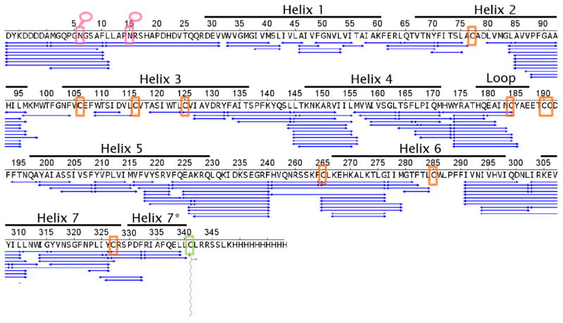
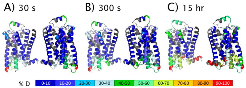
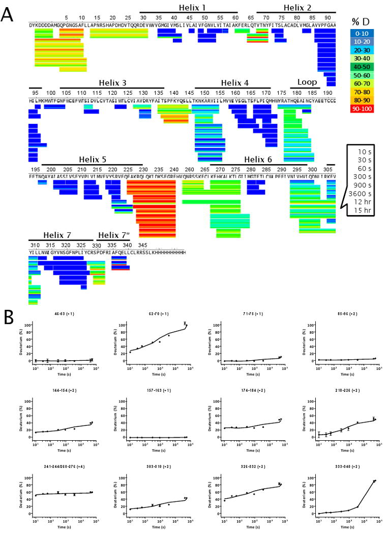
To examine the molecular details of ligand activation of G-protein coupled receptors (GPCRs), emphasis has been placed on structure determination of these receptors with stabilizing ligands. Here we present the methodology for receptor dynamics characterization of the GPCR human beta(2) adrenergic receptor bound to the inverse agonist carazolol using the technique of amide hydrogen/deuterium exchange coupled with mass spectrometry (HDX MS). The HDX MS profile of receptor bound to carazolol is consistent with thermal parameter observations in the crystal structure and provides additional information in highly dynamic regions of the receptor and chemical modifications demonstrating the highly complementary nature of the techniques. After optimization of HDX experimental conditions for this membrane protein, better than 89% sequence coverage was obtained for the receptor. The methodology presented paves the way for future analysis of beta(2)AR bound to pharmacologically distinct ligands as well as analysis of other GPCR family members.


Figures

Figure 1
Figure 1
Western blot analysis of carazolol-bound β2AR_460 after PNGase F incubation at 4°C for 1 hour, probed by using anti-FLAG (A), following separation by SDS-PAGE gel 10% Bis-Tris stained by SimplyBlue Safestain (B).
Figure 2
Figure 2
Peptide sequence map showing the MS/MS and MS verified coverage of carazolol-bound β2AR obtained under experimental conditions used in HDX experiments: 15 μM deglycosylated β2AR digested online using the immobilized pepsin column at room temperature (22 °C) and the quench solution containing 0.1 M NaPi_HCl (pH 2.4) 0.02% (m/v) DDM and 10-15 mM TCEP. Pink box dictates sites of N-glycosylation. Orange box indicates a cysteine residue (see Supplemental Table 1). Green box dictates site of protein palmitoylation.
Figure 3
Figure 3
HDX profile of carazolol-bound β2AR mapped to a model derived from the 3-D crystal structure of β2AR T4L (PDB: 2RH1) after exchange time of 30 s (A), 300 s (B) and 15 hr (C). Two views are represented for each timepoint. Each pair corresponds to rotation through 180°. The structure model was derived from 2RH1 with removal of T4L amino acids to reflect the construct used for HDX studies. All non-protein molecules were removed with the exception of carazolol. Gray cartoons represent the sequences not presented in the HDX profile.
Figure 4
Figure 4
HDX profile of deglycosylated β2AR mapped to its sequence after exchange time of 10 s, 30 s, 60 s, 300 s, 900 s, 3600 s, 12 h and 15 h and discarding the first two amino acid residues at the N-terminus of each peptide (A), and graphs of %D as a function of exchange time in log scale for selected peptide ions shown in panel A (B). The error bars in panel B were plotted as the standard deviation of four independent replicates, although in some cases the error bars are too small to be visible.

References

    1. Hanson MA, Stevens RC. Structure (Cambridge, MA, United States) 2009;17:8–14. - PubMed
    1. Kroeze WK, Sheffler DJ, Roth BL. Journal of Cell Science. 2003;116:4867–4869. - PubMed
    1. Conn PJ, Christopoulos A, Lindsley CW. Nature Reviews Drug Discovery. 2009;8:41–54. - PMC - PubMed
    1. Golan DE, T AH, Jr, Armstrong EJ, Armstrong AW. Principles of pharmacology: The pathophysiologic Basis of Drug Therapy. 2nd. Lippincott Williams & Wilkins; Baltimore, MD: 2008.
    1. Penn RB. Proceedings of the National Academy of Sciences. 2009;106:2095–2096. - PMC - PubMed

Publication types

MeSH terms