

Array lead zirconate titanate/glass piezoelectric microcantilevers for real-time detection of Bacillus anthracis with 10 spores/ml sensitivity and 1/1000 selectivity in bacterial mixtures
- PMID: 20059167
- PMCID: PMC2802521
- DOI: 10.1063/1.3264082
Array lead zirconate titanate/glass piezoelectric microcantilevers for real-time detection of Bacillus anthracis with 10 spores/ml sensitivity and 1/1000 selectivity in bacterial mixtures
Abstract
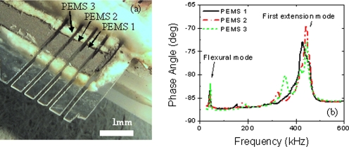
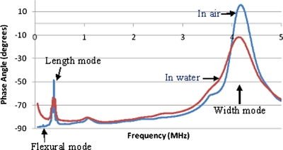
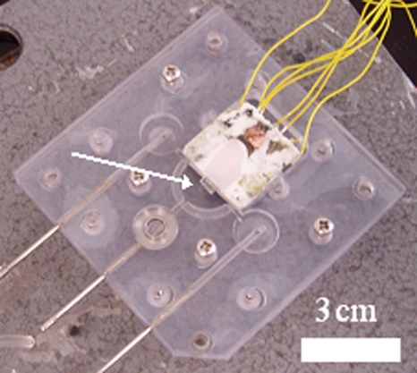
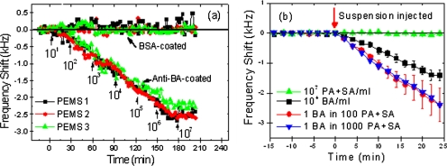
An array of three identical piezoelectric microcantilever sensors (PEMSs) consisting of a lead zirconate titanate layer bonded to a glass layer was fabricated and examined for simultaneous, in situ, real-time, all-electrical detection of Bacillus anthracis (BA) spores in an aqueous suspension using the first longitudinal extension mode of resonance. With anti-BA antibody immobilized on the sensor surfaces all three PEMS exhibited identical BA detection resonance frequency shifts at all tested concentrations, 10-10(7) spores/ml with a standard deviation of less than 10%. The detection concentration limit of 10 spores/ml was about two orders of magnitude lower than would be permitted by flexural peaks. In blinded-sample testing, the array PEMS detected BA in three samples containing BA: (1) 3.3x10(3) spores/ml, (2) a mixture of 3.3x10(3) spores/ml and 3.3x10(5) S. aureus (SA) and P. aeruginosa (PA) per ml, and (3) a mixture of 3.3x10(3) spores/ml with 3.3x10(6) SA+PA/ml. There was no response to a sample containing only 3.3x10(6) SA+PA/ml. These results illustrate the sensitivity, specificity, reusability, and reliability of array PEMS for in situ, real-time detection of BA spores.
Figures
Similar articles
-
Piezoelectric-excited millimeter-sized cantilever (PEMC) sensors detect Bacillus anthracis at 300 spores/mL.Biosens Bioelectron. 2006 Mar 15;21(9):1684-92. doi: 10.1016/j.bios.2005.08.001. Epub 2005 Sep 19. Biosens Bioelectron. 2006. PMID: 16169715
-
In situ detection of Bacillus anthracis spores using fully submersible, self-exciting, self-sensing PMN-PT/Sn piezoelectric microcantilevers.Analyst. 2007 Aug;132(8):777-83. doi: 10.1039/b704579d. Epub 2007 Jun 18. Analyst. 2007. PMID: 17646877
-
Label-free flow-enhanced specific detection of Bacillus anthracis using a piezoelectric microcantilever sensor.Analyst. 2008 May;133(5):649-54. doi: 10.1039/b715948j. Epub 2008 Feb 29. Analyst. 2008. PMID: 18427687 Free PMC article.
-
In situ, in-liquid, all-electrical detection of Salmonella typhimurium using lead titanate zirconate/gold-coated glass cantilevers at any dipping depth.Biosens Bioelectron. 2007 Jun 15;22(12):3132-8. doi: 10.1016/j.bios.2007.02.005. Epub 2007 Feb 20. Biosens Bioelectron. 2007. PMID: 17387007 Free PMC article.
-
Application of the real-time PCR for the detection of airborne microbial pathogens in reference to the anthrax spores.J Microbiol Methods. 2003 May;53(2):141-7. doi: 10.1016/s0167-7012(03)00019-8. J Microbiol Methods. 2003. PMID: 12654485 Review.
Cited by
-
In situ, amplification-free double-stranded mutation detection at 60 copies/ml with thousand-fold wild type in urine.Biosens Bioelectron. 2018 Nov 15;119:221-229. doi: 10.1016/j.bios.2018.07.062. Epub 2018 Jul 31. Biosens Bioelectron. 2018. PMID: 30142581 Free PMC article.
References
-
- Oshiro R. K., Method 1604, US EPA Office of Water (4303T), Washington, D.C., 2002.
-
- C. R.Kline, Jr., IEEE Engineering in Medicine and Biology 21, 43 (2002). - PubMed
Publication types
MeSH terms
Substances
Grants and funding
LinkOut - more resources
Full Text Sources