

Identification of a potential pharmacological sanctuary for HIV type 1 in a fraction of CD4(+) primary cells
- PMID: 20059395
- PMCID: PMC2858927
- DOI: 10.1089/aid.2009.0044
Identification of a potential pharmacological sanctuary for HIV type 1 in a fraction of CD4(+) primary cells
Abstract
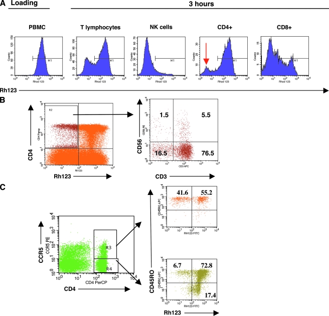
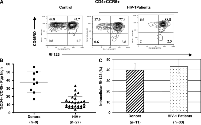
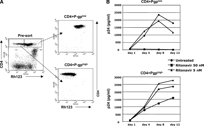
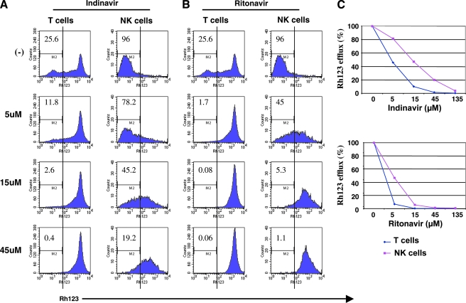
We have identified a subset of HIV-susceptible CD4(+)CCR5(+) cells in human PBMCs that can efficiently exclude protease inhibitors (PI) due to high P-glycoprotein (P-gp) efflux activity. Phenotypically these cells are heterogeneous, include both T and non-T cells, and some display markers of memory cells. Cells with high P-gp represent 16-56% (median = 37.3) of all CD4(+)CCR5(+) cells in healthy donors, and are selectively depleted in HIV-1-infected individuals (4.1-33%, median = 10.1). A fraction of primary cells productively infected by HIV-1, in vitro, have high P-gp pump activity, demonstrating that infection does not inhibit P-gp function. In agreement with these data, HIV-susceptible cells expressing high levels of P-gp require higher levels of PI for complete inhibition of virus spread. We conclude that the PI concentrations achieved in plasma could be suboptimal for full inhibition of virus spread in high P-gp cells, indicating that they may represent a pharmacological sanctuary for HIV-1.
Figures
Similar articles
-
HIV-protease inhibitors contribute to P-glycoprotein efflux function defect in peripheral blood lymphocytes from HIV-positive patients receiving HAART.J Acquir Immune Defic Syndr. 2001 Aug 1;27(4):321-30. doi: 10.1097/00126334-200108010-00001. J Acquir Immune Defic Syndr. 2001. PMID: 11468419
-
P-glycoprotein in blood CD4 cells of HIV-1-infected patients treated with protease inhibitors.HIV Med. 2003 Jan;4(1):67-71. doi: 10.1046/j.1468-1293.2003.00134.x. HIV Med. 2003. PMID: 12534962
-
P-Glycoprotein and transporter MRP1 reduce HIV protease inhibitor uptake in CD4 cells: potential for accelerated viral drug resistance?AIDS. 2001 Jul 27;15(11):1353-8. doi: 10.1097/00002030-200107270-00004. AIDS. 2001. PMID: 11504956
-
Transport of HIV protease inhibitors through the blood-brain barrier and interactions with the efflux proteins, P-glycoprotein and multidrug resistance proteins.J Acquir Immune Defic Syndr. 2004 Jun 1;36(2):649-58. doi: 10.1097/00126334-200406010-00001. J Acquir Immune Defic Syndr. 2004. PMID: 15167283 Review.
-
Combination of protease inhibitors for the treatment of HIV-1-infected patients: a review of pharmacokinetics and clinical experience.Antivir Ther. 2001 Dec;6(4):201-29. Antivir Ther. 2001. PMID: 11878403 Review.
Cited by
-
Analysis of multiple cell reservoirs expressing unspliced HIV-1 gag-pol mRNA in patients on antiretroviral therapy.Future Virol. 2012 Aug;7(8):819-832. doi: 10.2217/fvl.12.69. Future Virol. 2012. PMID: 23125871 Free PMC article.
-
High levels of CD2 expression identify HIV-1 latently infected resting memory CD4+ T cells in virally suppressed subjects.J Virol. 2013 Aug;87(16):9148-58. doi: 10.1128/JVI.01297-13. Epub 2013 Jun 12. J Virol. 2013. PMID: 23760244 Free PMC article.
-
P-glycoprotein (ABCB1) activity decreases raltegravir disposition in primary CD4+P-gphigh cells and correlates with HIV-1 viral load.J Antimicrob Chemother. 2016 Oct;71(10):2782-92. doi: 10.1093/jac/dkw215. Epub 2016 Jun 21. J Antimicrob Chemother. 2016. PMID: 27334660 Free PMC article.
-
Novel intravaginal nanomedicine for the targeted delivery of saquinavir to CD4+ immune cells.Int J Nanomedicine. 2013;8:2847-58. doi: 10.2147/IJN.S46958. Epub 2013 Aug 8. Int J Nanomedicine. 2013. PMID: 23966779 Free PMC article.
-
Varied sensitivity to therapy of HIV-1 strains in CD4+ lymphocyte sub-populations upon ART initiation.AIDS Res Ther. 2010 Dec 6;7:42. doi: 10.1186/1742-6405-7-42. AIDS Res Ther. 2010. PMID: 21134247 Free PMC article.
References
-
- Gulick RM. Mellors JW. Havlir D, et al. Treatment with indinavir, zidovudine, and lamivudine in adults with human immunodeficiency virus infection and prior antiretroviral therapy. N Engl J Med. 1997;337:734–739. - PubMed
-
- Ho DD. Neumann AU. Perelson AS. Chen W. Leonard JM. Markowitz M. Rapid turnover of plasma virions and CD4 lymphocytes in HIV-1 infection. Nature. 1995;373:123–126. - PubMed
-
- Perelson AS. Neumann AU. Markowitz M. Leonard JM. Ho DD. HIV-1 dynamics in vivo: Virion clearance rate, infected cell life-span, and viral generation time. Science. 1996;271:1582–1586. - PubMed
-
- Perelson AS. Essunger P. Cao Y, et al. Decay characteristics of HIV-1-infected compartments during combination therapy. Nature. 1997;387:188–191. - PubMed
-
- Wei X. Ghosh SK. Taylor ME, et al. Viral dynamics in human immunodeficiency virus type 1 infection. Nature. 1995;373:117–122. - PubMed
Publication types
MeSH terms
Substances
Grants and funding
LinkOut - more resources
Full Text Sources
Medical
Research Materials
Miscellaneous
