. 2010 May;130(1):114-24.
doi: 10.1111/j.1365-2567.2009.03218.x. Epub 2010 Jan 6.

Prevention and treatment of experimental autoimmune encephalomyelitis with clonotypic CDR3 peptides: CD4(+) Foxp3(+) T-regulatory cells suppress interleukin-2-dependent expansion of myelin basic protein-specific T cells

Affiliations

Prevention and treatment of experimental autoimmune encephalomyelitis with clonotypic CDR3 peptides: CD4(+) Foxp3(+) T-regulatory cells suppress interleukin-2-dependent expansion of myelin basic protein-specific T cells

Abigail C Buenafe et al. Immunology. 2010 May.

Abstract

T-cell receptor (TCR)-derived peptides are recognized by the immune system and are capable of modulating autoimmune responses. Using the myelin basic protein (MBP) TCR 1501 transgenic mouse model, we demonstrated that TCR CDR3 peptides from the transgenic TCR can provide a protective effect when therapy is initiated before the induction of experimental autoimmune encephalomyelitis (EAE). More importantly, TCR CDR3 peptide therapy can ameliorate the disease when administered after EAE onset. Concurrent with the therapeutic effects, we observed reduced T-cell proliferation and reduced interleukin-2 (IL-2) levels in response to stimulation with MBP-85-99 peptide in splenocyte cultures from mice receiving TCR CDR3 peptides compared with that of control mice. Moreover, we found that Foxp3(+) CD4 T cells from mice protected with TCR CDR3 peptide are preferentially expanded in the presence of IL-2. This is supportive of a proposed mechanism where Foxp3(+) T-regulatory cells induced by therapy with MBP-85-99 TCR CDR3 peptides limit expansion and the encephalitogenic activity of MBP-85-99-specific T cells by regulating the levels of secreted IL-2.


Figures
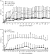
Figure 1
Figure 1
Myelin basic protein (MBP) 1501 transgenic mice induced for experimental autoimmune encephalomyelitis (EAE) are protected by T-cell receptor (TCR) CDR3 peptide/incomplete Freund’s adjuvant (IFA) but not by the IFA control. (a) TCR CDR3α peptide or TCR CDR3β peptide was injected intraperitoneally (i.p.) in IFA on days −7 (100 μg) and +3 (50 μg) relative to immunization with MBP-85-99/complete Freund’s adjuvant (CFA) (n = 9 or 10 mice per group). Mice also received weekly boosts of either TCR CDR3α peptide or TCR CDR3β peptide (all at 25 μg) beginning on day +10. Statistically significant differences from the IFA control group were determined using the Mann–Whitney test and were achieved in the TCR CDR3α peptide-treated group on days 15–20 and 34–42 (P ≤ 0·05). Significant differences were achieved in the TCR CDR3β peptide-treated group on days 15–22 and 32–42 (P ≤ 0·05). Cumulative disease score for each group: IFA control 87 ± 38; TCR CDR3α 41 ± 60 (P = 0·06 compared with the IFA control); TCR CDR3β 35 ± 39 (P = 0·01 compared with the IFA control). This experiment is also listed as Exp. 2 in Table 1. (b) Disease induction and the protective regimen were identical to that in (a) except that mice in the experimental group received both TCR CDR3α and TCR CDR3β peptides simultaneously. Disease score differences between the experimental and control groups were significant from day 12 to day 33 (P ≤ 0·002). Cumulative disease score for each group: IFA control 76 ± 20 (n = 7); TCR CDR3 peptides 13 ± 15 (n = 6; P = 0·00006 compared with the IFA control). Arrows indicate days when TCR peptide injection or control injection was given. This experiment is also listed as Exp. 3 in Table 1.
Figure 2
Figure 2
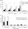
Proliferation to myelin basic protein (MBP)-85-99 peptide is suppressed in MBP 1501 transgenic mice protected with T-cell receptor (TCR) CDR3 peptides. (a) Spleens were pooled on day 42 after immunization with MBP-85-99/complete Freund’s adjuvant (CFA) from mice receiving TCR CDR3α (n = 5), TCR CDR3β (n = 5), or incomplete Freund’s adjuvant (IFA) (n = 4) as a protective regimen. Proliferative responses were assessed against MBP-85-99 (10, 2, or 0·4 μg/ml), concanavalin A (ConA) or medium alone. (b) Spleens were pooled on day 20 after immunization with MBP-85-99/CFA from mice receiving TCR CDR3α + TCR CDR3β (n = 9) or control TCR CDR2α + TCR CDR2β peptides (n = 9) as a protective regimen. Proliferative responses were assessed against 50 or 10 μg/ml of MBP-85-99, TCR CDR3 peptides, control TCR CDR2 peptides, or medium. *P < 0·005; #P = 0·018.
Figure 3
Figure 3
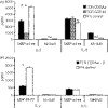
Interleukin (IL)-2 levels are suppressed in response to myelin basic protein (MBP)-85-99 in MBP 1501 transgenic mice protected with T-cell receptor (TCR) CDR3 peptides. (a) Splenocytes isolated from mice protected with TCR CDR3α, TCR CDR3β, or incomplete Freund’s adjuvant (IFA) on day 42 or (b) from mice protected with TCR CDR3α + TCR CDR3β peptides or IFA on day 33 after immunization were stimulated with MBP-85-99 or medium. Culture supernatants were collected after 72 hr and assessed for cytokine production using the Luminex assay. IL-2 levels were significantly lower in TCR CDR3-protected mice, but no differences in IL-6 levels were observed. *P ≤ 0·002; #P = 0·005; nd, not done.
Figure 4
Figure 4
Protective treatment with T-cell receptor (TCR) CDR3α + CDR3β peptides results in enhanced expression of FoxP3 in CD4+ splenocytes. (a) Spleen cells were obtained from myelin basic protein (MBP) 1501 transgenic mice induced for experimental autoimmune encephalomyelitis (EAE) and protected by TCR CDR3α + TCR CDR3β peptides, or by irrelevant TCR CDR2 peptides as a control. Spleens were pooled (n = 9 per group) and analyzed by flow cytometry for surface CD4 and intracellular FoxP3 expression. (b) In a separate experiment, spleen cells were pooled from four MBP 1501 transgenic mice induced for EAE and protected by TCR CDR3α + TCR CDR3β peptides or from four incomplete Freund's adjuvant (IFA) controls, then labelled with carboxyfluorescein diacetate succinimyl ester (CFSE) and cultured for 5 days with or without IL-2. IL-2 induced greater proliferation of FoxP3+ T cells in the TCR CDR3 peptide group compared with the control group (57% versus 29%, calculated by dividing the top left quadrant by the top left + top right quadrants).
Figure 6
Figure 6
Interleukin (IL)-2 levels are suppressed in response to myelin basic protein (MBP)-85-99 in MBP 1501 transgenic mice treated with T-cell receptor (TCR) CDR3 peptides. Active experimental autoimmune encephalomyelitis (EAE) was induced in MBP 1501 mice by immunization with MBP-85-99 peptide/complete Freund’s adjuvant (CFA) and, at disease onset, treated with TCR CDR3α + TCR CDR3β peptides or with irrelevant TCR CDR2α + TCR CDR2β peptides. After four injections of peptide given at weekly intervals, pooled splenocytes were stimulated with MBP-85-99, TCR CDR3 peptides, control TCR CDR2 peptides, or medium. Culture supernatants were collected after 72 hr and assessed for IL-2 production by enzyme-linked immunosorbent assay (ELISA) analysis. *P = 0·014.
Figure 5
Figure 5
T-cell receptor (TCR) CDR3 peptides treat ongoing experimental autoimmune encephalomyelitis (EAE) in myelin basic protein (MBP) 1501 transgenic mice. Active EAE was induced in MBP 1501 mice by immunization with 200 μg of MBP-85-99 peptide in complete Freund’s adjuvant (CFA) with 400 μg of Mycobacterium tuberculosis. Upon disease onset at a score of 1·5 or 2·0, mice received injections of 100 μg of each of TCR CDR3α + TCR CDR3β peptides subcutaneously in incomplete Freund’s adjuvant (IFA). Control mice received 100 μg each of irrelevant TCR CDR2α + TCR CDR2β peptides. TCR peptide injections were repeated weekly at the same dose. Significant differences between treated and control groups were achieved from day 10 to day 32 (P < 0·03). Arrows indicate days when TCR peptide injection was given. Disease course data are presented under Exp. 1 in Table 2.

References

    1. Olerup O, Hillert J. HLA class II-associated genetic susceptibility to multiple sclerosis: a critical evaluation. Tissue Antigens. 1991;38:1–15. - PubMed
    1. Haines JL, Ter-Minassian M, Bazyk A, et al. A complete genomic screen for multiple sclerosis underscores a role for the major histocompatibility complex. The Multiple Sclerosis Genetics Group. Nat Genet. 1996;13:469–71. - PubMed
    1. Sawcer S, Jones HB, Feakes R, et al. A genome screen in multiple sclerosis reveals susceptibility loci on chromosome 6p21 and 17q22. Nat Genet. 1996;13:464–8. - PubMed
    1. Ebers GC, Kukay K, Bulman DE, et al. A full genome search in multiple sclerosis. Nat Genet. 1996;13:472–6. - PubMed
    1. Hauser SL, Fleischnick E, Weiner HL, Marcus D, Awdeh Z, Yunis EJ, Alper CA. Extended major histocompatibility haplotypes in patients with multiple sclerosis. Neurology. 1989;39:275–7. - PubMed

Publication types

MeSH terms