Review
. 2010 Jan;267(1):71-88.
doi: 10.1111/j.1365-2796.2009.02184.x.

Emerging peptide nanomedicine to regenerate tissues and organs

Affiliations
Review

Emerging peptide nanomedicine to regenerate tissues and organs

M J Webber et al. J Intern Med. 2010 Jan.

Abstract

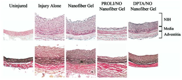
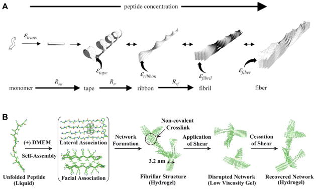
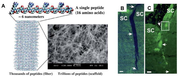
Peptide nanostructures containing bioactive signals offer exciting novel therapies of broad potential impact in regenerative medicine. These nanostructures can be designed through self-assembly strategies and supramolecular chemistry, and have the potential to combine bioactivity for multiple targets with biocompatibility. It is also possible to multiplex their functions by using them to deliver proteins, nucleic acids, drugs and cells. In this review, we illustrate progress made in this new field by our group and others using peptide-based nanotechnology. Specifically, we highlight the use of self-assembling peptide amphiphiles towards applications in the regeneration of the central nervous system, vasculature and hard tissue along with the transplant of islets and the controlled release of nitric oxide to prevent neointimal hyperplasia. Also, we discuss other self-assembling oligopeptide technology and the progress made with these materials towards the development of potential therapies.


Figures

Figure 1
Figure 1
(A) Molecular Structure of a representative peptide amphiphile with four rationally designed chemical entities. (B) Molecular graphics illustration of an IKVAV-containing peptide amphiphile molecule and its self-assembly into nanofibers; (C) TEM micrograph of IKVAV nanofibers; (D) SEM micrograph of IKVAV nanofiber gel network. Reprinted with permission from Gabriel Silva et. al., Selective differentiation of neural progenitor cells by high-epitope density nanofibers, Science, 2004; 303, 1352–1355 (parts B, D) and with permission from Krista Niece et. al., Self-assembly combining two bioactive peptide-amphiphile molecules into nanofibers by electrostatic attraction, Journal of the American Chemical Society, 2003; 125(24), 7146–7147 (part C).
Figure 2
Figure 2
(A) Therapeutic bone marrow mononuclear cells derived from a luciferase transgene mouse and transplanted within an RGDS-presenting PA network into either flank of a wild-type mouse, imaged at 4 days following transplant; (B) The same cells when transplanted within a PA network that did not contain the RGDS eptiope; (C) The same cells, transplanted with only a saline vehicle. Reprinted with permission from Matthew Webber et. al., Development of bioactive peptide amphiphiles for therapeutic cell delivery, Acta Biomaterialia, 2009; in press.
Figure 3
Figure 3
(A) Schematic representation of one method to form a self-sealing closed sac. A sample of the denser negatively charged biopolymer solution is dropped onto a positively charged peptide amphiphile (PA) solution. (B) Open and (C) closed sac formed by injection of a fluorescently tagged hyaluronic acid (HA) solution into a PA solution. (D) Self-assembled sacs of varying sizes. (E) PA-HA membranes of different shapes created by interfacing the large- and small-molecule solutions in a very shallow template ( ~1 mm thick). (F) Continuous strings pulled from the interface between the PA and HA solutions. Reprinted with permission from Ramille Capito et. al., Self-assembly of large and small molecules into hierarchically ordered sacs and membranes, Science, 2008; 319, 1812–1816.
Figure 4
Figure 4
IKVAV PA promotes regeneration of descending corticospinal motor axons after spinal cord injury. a, b: Representative Neurolucida tracings of BDA-labeled descending motor fibers within a distance of 500 μm rostral of the lesion in vehicle-injected (a) and IKVAV PA-injected (b) animals. The dotted lines demarcate the borders of the lesion. c–f: Bright-field images of BDA-labeled tracts in lesion (c, e) and caudal to lesion (d, f) used for Neurolucida tracings in an IKVAV PA-injected spinal cord (a, b). g, h: Bar graphs show the extent to which labeled corticospinal axons penetrated the lesion. *The groups representing three control and three IKVAV PA mice and the tracing of 130 individual axons differ from each other at p < 0.03 by the Wilcoxon rank test. (R, Rostral; C, caudal; D, dorsal; V, ventral. Scale bars: a–d: 100 μm and e,f: 25 μm.) Reprinted with permission from Vicki Tysseling-Mattiace et. al., Self-assembling nanofibers inhibit glial scar formation and promote axon elongation after spinal cord injury, Journal of Neuroscience, 2008; 28(14), 3814–3823.
Figure 5
Figure 5
In vivo angiogenesis assay. Rat cornea photographs 10 days after the placement of various materials at the site indicated by the black arrow. (A)Heparin-nucleated PA nanofiber networks with growth factors show extensive neovascularization. Controls of collagen, heparin, and growth factors (B) and collagen with growth factors (C) show some neovascularization. Heparin with growth factors (D), and collagen with heparin. The bar graph (F) contains values for the average and maximum length of new blood vessels and the area of corneal neovascularization. A 100% value in the area measurement indicates that the cornea is completely covered, and a 100% value in the length parameters indicates that the new vessels are as long as the diameter of the cornea (bars are 95% confidence levels, * p < 0.05 when PA-heparin gel was compared to collagen gel with growth factors, ** p < 0.005 when PA-heparin gel with growth factors was compared to all of the other controls). PA nanofibers with heparin, PA solution with growth factors, and growth factors alone did not result in measurable neovascularization (values not shown in graph). Reprinted with permission from Kanya Rajangam et. al., Heparin binding nanostructures to promote the growth of blood vessels, Nano Letters, 2006; 6(9), 2086–2090.
Figure 6
Figure 6
Rat carotid artery sections from uninjured, injury alone, nanofiber gel, PROLI/NO nanofiber gel, and DPTA/NO nanofiber gel animals euthanized at 14 days (n=6 per group). Displayed are representative sections (100x original magnification)from each group using routine staining with hematoxylin and eosin (top) and Verhoff-van Gieson (bottom). PROLI/NO and DPTA/NO are two small molecule diazeniumdiolate nitric oxide donors that were mixed with the PA nanofiber gel. Reprinted with permission from Muneera Kapadia et. al., Nitric oxide and nanotechnology: A novel approach to inhibit neointimal hyperplasia, Journal of Vascular Surgery, 2008 47(1), 173–182.
Figure 7
Figure 7
(A) The hierarchical equilibrium configurations of self-assembly of β-sheet forming peptides, which assemble to form tapes, ribbons, fibrils and fibers depending on parameters such as peptide concentration, solution pH, or sequence/charge of the precursor monomer. Reprinted with permission from Amalia Aggeli et. al., pH as a Trigger of Peptide β-sheet Self-Assembly and Reversible Switching between Nematic Iotropic Phases, Journal of the American Chemical Society, 2003 125(3), 9619–9628. (B) The self-assembly of β-hairpin peptides through charge screening in physiologic media (DMEM) allows for the formation of self-healing hydrogels that can be syringe-delivered. Reprinted from Lisa Haines-Butterick et. al., Controlling hydrogelation kinetics by peptide design for three-dimensional encapsulation and injectable delivery of cells, 2007 104(19), 7791–7796 Copyright 2007 National Academy of Sciences USA.
Figure 8
Figure 8
(A) Ionic self-complimentary peptides self-assemble by hydrogen bonding into fibers consisting of thousands of peptides which further entangle into scaffolds that are ~99.5% water and ~0.5% peptide. Reprinted with permission from Fabrizio Gelain et. al., Designer self-assembling peptide scaffolds for 3-d tissue cell cultures and regenerative medicine, Macromolecular Bioscience, 2007 7(5), 544–551. (B, C) View of the superior colliculus (SC) 30 days following the formation of a tissue gap by deep transection of the optic tract in the hamster midbrain following a control treatment with saline (B) or treatment with ionic self-complimentary peptide nanofibers (C). With nanofiber treatment, the site of the lesion has healed and axons have grown through the treated area and reached the caudal part of the SC. Reprinted from Rutledge G. Ellis-Behnke et. al., Nano neuro knitting: Peptide nanofiber scaffold for brain repair and axon regeneration with functional return of vision,, 2006 103(13), 5054–5059 Copyright 2006 National Academy of Sciences USA.

References

    1. Gurtner GC, Callaghan MJ, Longaker MT. Progress and potential for regenerative medicine. Annu Rev Med. 2007;58:299–312. - PubMed
    1. Hartgerink JD, Beniash E, Stupp SI. Self-assembly and mineralization of peptide-amphiphile nanofibers. Science. 2001;294:1684–8. - PubMed
    1. Harrison BS, Atala A. Carbon nanotube applications for tissue engineering. Biomaterials. 2007;28:344–53. - PubMed
    1. Tran PA, Zhang L, Webster TJ. Carbon nanofibers and carbon nanotubes in regenerative medicine. Adv Drug Deliv Rev. 2009 - PubMed
    1. Jamieson T, Bakhshi R, Petrova D, Pocock R, Imani M, Seifalian AM. Biological applications of quantum dots. Biomaterials. 2007;28:4717–32. - PubMed