. 2010 May;22(5):e138-51.
doi: 10.1111/j.1365-2982.2009.01448.x. Epub 2010 Jan 5.

ICC-MY coordinate smooth muscle electrical and mechanical activity in the murine small intestine

Affiliations

ICC-MY coordinate smooth muscle electrical and mechanical activity in the murine small intestine

G W Hennig et al. Neurogastroenterol Motil. 2010 May.

Abstract

Background: Animals carrying genetic mutations have provided powerful insights into the role of interstitial cells of Cajal (ICC) in motility. One classic model is the W/W(V) mouse which carries loss-of-function mutations in c-kit alleles, but retains minimal function of the tyrosine kinase. Previous studies have documented loss of slow waves and aberrant motility in the small intestine of W/W(V) mice where myenteric ICC (ICC-MY) are significantly depleted.

Methods: Here, we used morphological and electrophysiological techniques to further assess the loss of ICC around the circumference of the small intestine and determine consequences of losing ICC-MY on electrical activity, Ca(2+) transients and contractions of the longitudinal muscle (LM).

Key results: In wild-type mice, there was coherent propagation of Ca(2+) transients through the ICC-MY network and spread of this activity to the LM. In short segments of small intestine in vitro and in exteriorized segments, slow waves coordinated smoothly propagating Ca(2+) waves and contractions in the LM of wild-type mice. In W/W(V) mice, Ca(2+) waves were initiated at variable sites along and around intestinal segments and propagated without constraint unless they collided with other Ca(2+) waves. This activity resulted in abrupt, uncoordinated contractions.

Conclusions & inferences: These results show how dominance of pacemaking by ICC-MY coordinates propagating con-tractions and regulates the spontaneous activity of smooth muscle.


Conflict of interest statement

Competing Interests: the authors have no competing interests.

Figures

Figure 1
Figure 1
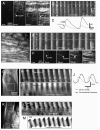
Preparations used to study Ca2+ transients in ICC-MY and LM and longitudinal contractions in the mouse small intestine from the cellular to organ level. A & B. show a flat-sheet preparation used to examine the spread of Ca2+ transients at the cellular level where single LM cells and underlying ICC-MY could be resolved. In some experiments the LM was removed to better resolve the activity of ICC-MY. To examine the spread of Ca2+ waves in the longitudinal axis and around the circumference of the small bowel, isolated tubular preparations (D) or exteriorized loops of small intestine (E) were used. LM contractions were monitored using a grid of surface markers positioned on flat-sheet preparations (F&G). The distance between pairs of markers were calculated dynamically, expressed in grayscale (see vertical bars in G), and were used to create spatio-temporal maps (H) showing the change in LM contraction (I) over time. Spatio-temporal cubes were constructed by constructing a grid of quadrangles between surface markers (J). Each corner of each quadrangle was positioned midway between relevant pairs of surface markers (J (i)). The color at each corner corresponded to the longitudinal distance between relevant pairs of surface markers. Individual quadrangles were filled by blending the colors at each corner throughout the quadrangle using linear interpolation (J (ii)). Pixels that were above a certain level of contraction (color) were thresholded (J (iii)) and a marching cubes algorithm was used to construct 3 dimensional spatio-temporal objects (J (iv)) from thresholded areas in frames throughout the movie.
Figure 2
Figure 2
ICC networks and electrical slow waves in the small intestines of W/WV mutants. A dense network of ICC-MY (see arrowheads) and ICC-DMP (see arrows) was observed from the mesenteric to the anti-mesenteric region of wild-type mouse small intestine (A–C). Slow waves with similar characteristics were recorded at each of these positions (below anatomical panels). In W/WV mice isolated patches of ICC-MY (arrowheads) were observed in the mesenteric region (D), and, in some cases, midway between the mesenteric and anti-mesenteric regions (E). ICC-MY were rare in the anti-mesenteric region (F). ICC-DMP (arrows) were unaffected in all three regions, as previously reported . In a few impalements in the mesenteric region, very low amplitude slow waves were recorded (D; trace below anatomical panel). Little or no rhythmic slow wave activity was recorded at other positions around the circumference of the small intestine. Scale bars in panels C and F apply to panels A–C and D–F respectively.
Figure 3
Figure 3
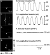
Electrical activity recorded from the circular (CM) and longitudinal muscle (LM) cells of wild-type (+/+) and W/WV mutants. CM and LM displayed similar slow wave activity in wild-type small intestinal muscles (A–D). Panels A and B show a propridium iodide (PI) filled CM cell and slow wave activity recorded from the cell that was filled. Panels C and D show PI filling of a LM cell and slow wave activity recorded from the cell. Slow wave activity was not observed in the CM and LM of W/WV muscles. Panels E and F show PI filled cell from the CM lacking slow wave activity and panels G and H show a PI filled cell from the LM also lacking slow wave activity. Impaled smooth muscles were in the anti-mesenteric region. All recordings were taken in the presence of nifedipine, 1M, to stabilize the muscles during filling. Scale bars are as indicated in each panel. Directions of CM and LM are indicated adjacent to panel G.
Figure 4
Figure 4
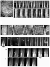
Spread of Ca2+ transients and LM contractions in wild-type small intestine. In a preparation in which the LM was partially removed, Ca2+transients spread in the ICC-MY network (A). The frequency and direction of propagation of Ca2+ transients in ICC-MY and LM were consistent throughout recording periods (B). Ca2+ increased approximately 0.1s before LM fibers (running horizontally) overlying the ICC-MY network were activated, indicating ICC-MY pace LM (C). Ca2+ waves with consistent frequency and direction were also observed in an preparation with intact LM (D&E), Faint Ca2+ transients were observed in ICC-MY approximately 0.1s before LM was activated (F). In isolated tubular preparations (G), the frequency, velocity and direction of propagating Ca2+ waves was constant during periods of recording (H). Ca2+ transients in the LM preceded circumferential contraction (I) by approximately 0.5–1s (J). In exteriorized loops (K) a similar constant patterns of Ca2+ waves were observed (L) prior to longitudinal contractions (M).
Figure 5
Figure 5
Spread of Ca2+ transients and LM contraction in W/WV small intestine. In flat-sheet preparations (A) the circumferential spread of Ca2+ transients in LM was irregular with dramatic changes in frequency, direction and length of propagation (see asterisks in B & C). In some preparations trains of Ca2+ transients were observed in LM (C). A disordered pattern of Ca2+ transients was also observed in isolated tubular preparations (D & E), and resulted irregular, abrupt movements (E). Ca2+ transients in the LM often propagated for at least several millimeters at high velocity (F–I), however the site of initiation was variable and LM transients were not coordinated around the circumference of the small intestine.
Figure 6
Figure 6
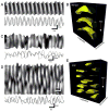
Pattern of LM contraction in wild-type and W/WV mice. LM contractions were constant in wild-type mice during recording periods, with contractions originating at the same frequency from the same region of the preparation and propagated in the aboral direction at constant velocities (A). The consistent LM contractions around the circumference of the small bowel are portrayed in the spatio-temporal cubes in B. In W/WV mice, LM contractions were abrupt and originated at variable positions within the preparations (C). Multiple initiation sites were observed in the small intestine of W/WV mice and propagating events collided (D & E).
Figure 7
Figure 7
Effects of block of L-type Ca2+ channels and depolarization. Contractions in W/WV mice (A) were reliant upon Ca2+ entry through L-Type Ca2+ channels because nicardipine (2μM) blocked all contractions (B). Propagating contractions in wild-type small intestines (C) were disrupted by elevation of external K+ (to 10 mM: D) and the number of pacemaker initiation sites increased. Higher concentrations of external K+ (30 mM: E) compounded this effect, however the amplitude of contractions decreased.

Similar articles

Cited by

References

    1. Vanderwinden JM, Rumessen JJ. Interstitial cells of Cajal in human gut and gastrointestinal disease. Microsc Res Tech. 1999;47:344–360. - PubMed
    1. Sanders KM, Ordög T, Koh SD, Torihashi S, Ward SM. Development and plasticity of interstitial cells of Cajal. Neurogastroenterol Motil. 1999;11:311–38. - PubMed
    1. Burns AJ. Disorders of interstitial cells of Cajal. J Pediatr Gastroenterol Nutr. 2007;45:103–6. - PubMed
    1. Rolle U, Piaseczna-Piotrowska A, Puri P. Interstitial cells of Cajal in the normal gut and in intestinal motility disorders of childhood. Pediatr Surg Int. 2007;23:1139–52. - PubMed
    1. Farrugia G. Interstitial cells of Cajal in health and disease. Neurogastroenterol Motil. 2008;20:54–63. - PubMed

Publication types