. 2009 Dec;17(6):861-73.
doi: 10.1016/j.devcel.2009.10.010.

Bone ridge patterning during musculoskeletal assembly is mediated through SCX regulation of Bmp4 at the tendon-skeleton junction

Affiliations

Bone ridge patterning during musculoskeletal assembly is mediated through SCX regulation of Bmp4 at the tendon-skeleton junction

Einat Blitz et al. Dev Cell. 2009 Dec.

Abstract

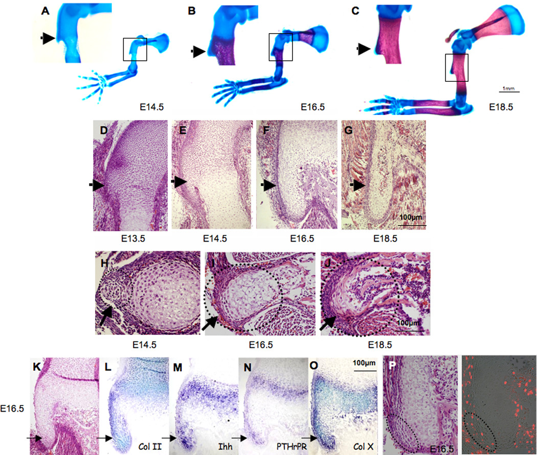
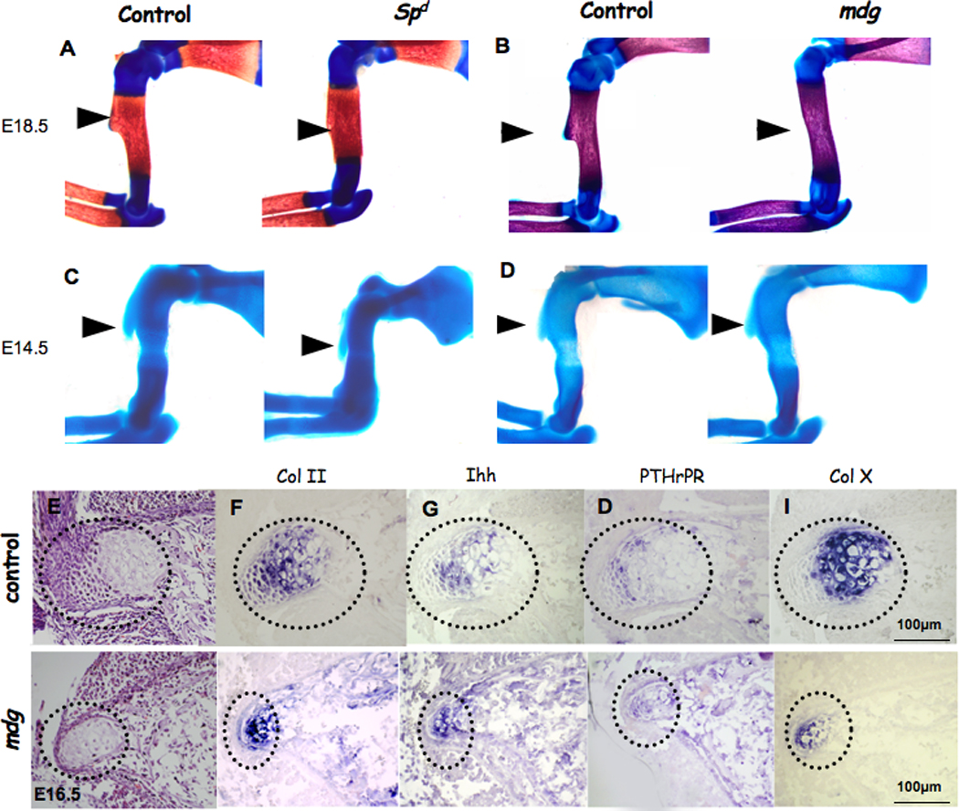
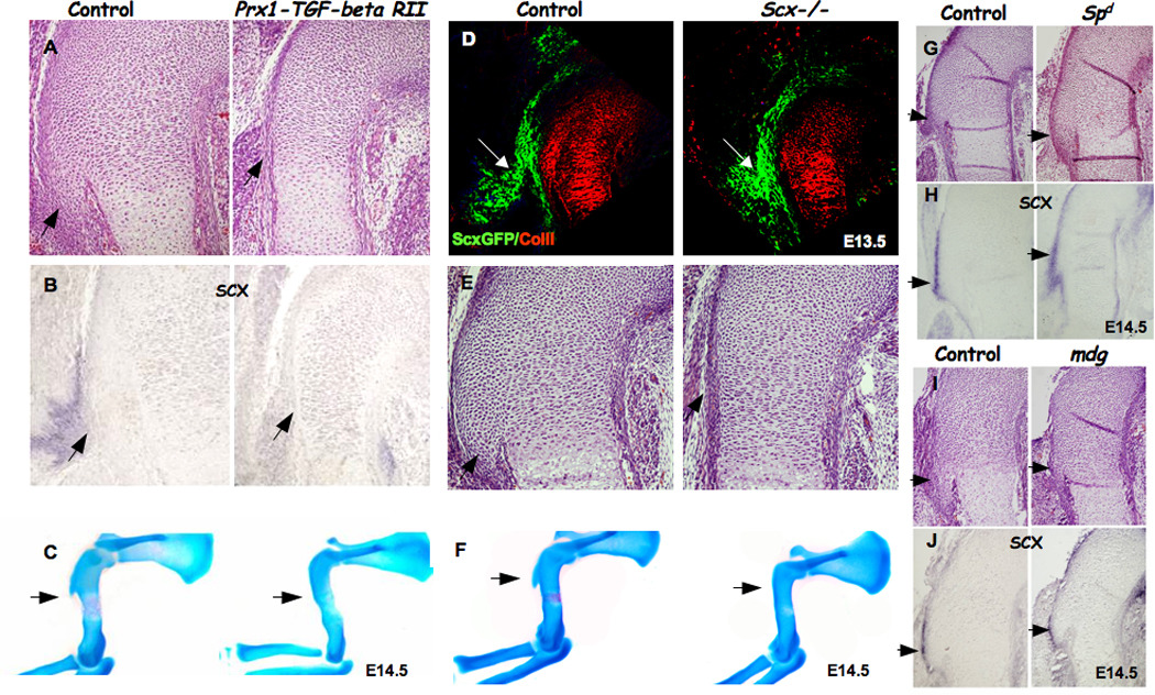
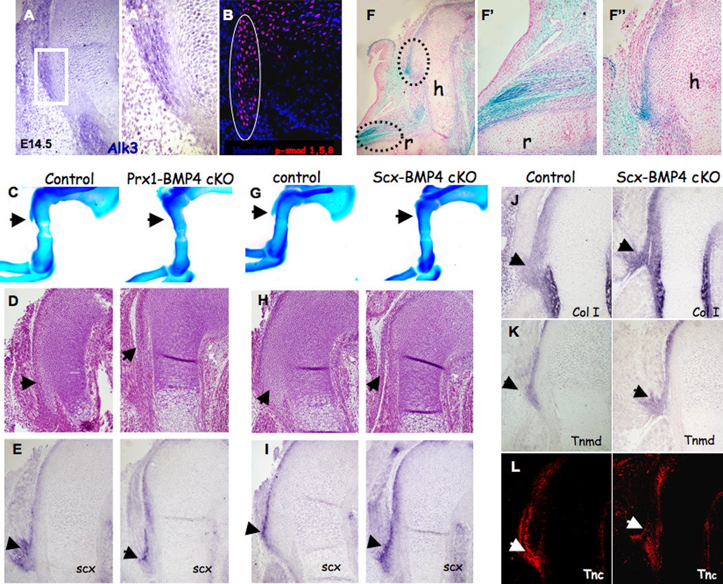
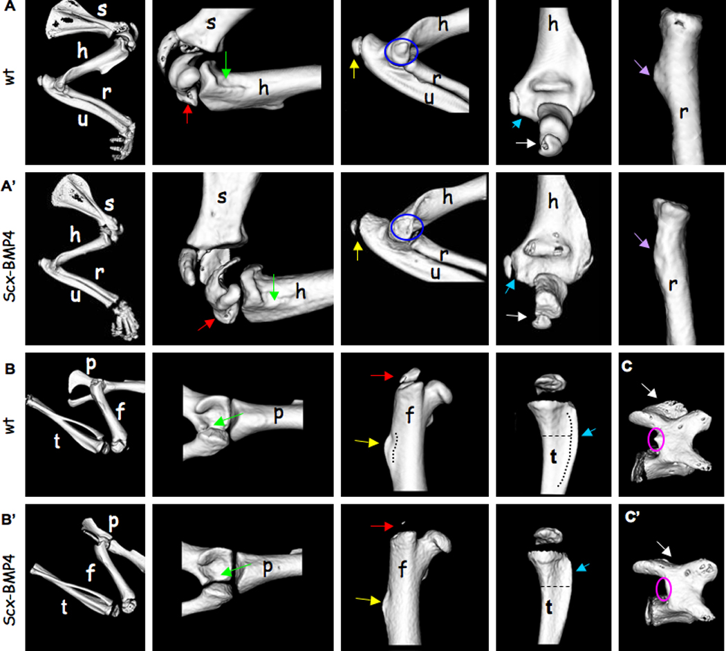
During the assembly of the musculoskeletal system, bone ridges provide a stable anchoring point and stress dissipation for the attachment of muscles via tendons to the skeleton. In this study, we investigate the development of the deltoid tuberosity as a model for bone ridge formation. We show that the deltoid tuberosity develops through endochondral ossification in a two-phase process: initiation is regulated by a signal from the tendons, whereas the subsequent growth phase is muscle dependent. We then show that the transcription factor scleraxis (SCX) regulates Bmp4 in tendon cells at their insertion site. The inhibition of deltoid tuberosity formation and several other bone ridges in embryos in which Bmp4 expression was blocked specifically in Scx-expressing cells implicates BMP4 as a key mediator of tendon effects on bone ridge formation. This study establishes a mechanistic basis for tendon-skeleton regulatory interactions during musculoskeletal assembly and bone secondary patterning.


Figures

Figure 1
Figure 1. Developmental analysis of the deltoid tuberosity
(A–C) Skeletal preparations of E14.5, E16.5 and E18.5 WT embryos (the boxed areas of the DT are enlarged on the left). (D–J) Histological sagittal (D–G) and transverse (H–J) sections through the DT of E13.5–E18.5 WT mice (dotted circles indicate DT area). (K–O) Tuberosity cell differentiation is demonstrated by the expression of growth plate markers. (K) Histological sagittal section through the DT of E16.5 WT mice. (L–O) In situ hybridization analysis of DT sagittal sections from E16.5 WT mice, using antisense complementary RNA probes for collagen II (Col II), Indian hedgehog (Ihh), parathyroid hormone-related peptide receptor (PTHrPR) and collagen X (Col X) mRNA. (P) BrdU incorporation in E16.5 WT mice; dotted circles indicate tuberosity tip. (Throughout, black arrows indicate DT.)
Figure 2
Figure 2. Bone ridge development is disrupted in mice with defective musculature
(A–D) Forelimbs of E18.5 (A–B) and E14.5 (C–D) control, Spd/Spd and mdg/mdg mice stained with Alcian blue and Alizarin red (throughout, black arrows indicate DT). (E–I) Expression of growth plate markers in paralyzed embryos (dotted circles indicate DT area). (E) Histological transverse sections of control and mdg mutant E16.5 tuberosity. (F–I) In situ hybridization analysis of DT transverse sections from E16.5 control and mdg mice using antisense complementary RNA probes for collagen II (Col II), Indian hedgehog (Ihh), parathyroid hormone-related peptide receptor (PTHrPR) and collagen X (Col X) mRNA.
Figure 3
Figure 3. Tendons regulate deltoid tuberosity initiation through scleraxis
(A–B) Detection of scleraxis expression as an indication for tendon formation in Prx1-Tgf-βRII embryos. (A) Histological sagittal sections through the humeri of control and Prx1-Tgf-βRII mice. (B) In situ hybridization of sections through the humeri of control and Prx1-Tgf-βRII embryos at E14.5, using Scx probe. (C) skeleton preparation of control and Prx1-Tgf-βRII mice. (D) Sagittal sections through the humerus of control and Scx−/− embryos carrying the ScxGFP reporter and counterstained with anti-collagen II antibody. White arrows mark tendon attachment site and the presumable initiation site of the DT (green: tendon, red: cartilage). (E–F) Detection of DT by (E) histological sagittal sections through the humerus and (F) skeleton preparation of E14.5 Scx−/− mutant. (G–J) Scx expression in control, muscle-less (homozygous Spd) and paralyzed (mdg) mice. (G,I) Histological sagittal sections through the humerus of control, Spd and mdg mutant mice. (H,J) In situ hybridization through the humeri of control and Spd and mdg embryos at E14.5, using Scx probe (throughout, black arrows point at DT).
Figure 4
Figure 4. BMP4 acts downstream of scleraxis
(A) Double Fluorescent in situ hybridization of DT sagittal sections from E13.5 WT mice, using antisense complementary RNA probes for Scx and Bmp4; white arrows mark tendon insertion site. (B) In situ hybridization of forelimb joint sagittal sections from E14.5 WT mice, using antisense complementary RNA probes for Scx and Bmp4. (C–D) In situ hybridization analysis of sagittal sections of humerus at E13.5, using antisense complementary RNA probes for Scx and Bmp4; black arrows mark tendon insertion site. (E–F) qRT-PCR analysis of Bmp4 mRNA levels in C3T1/2H10 cells transiently (E) or stably (F) transfected with Scx. Stable cells (F) were transfected with empty vector or Scx and passed selection or kept in culture without transfection (n/t). (G) Luciferase activity in HeLa cells transfected with indicated plasmids: PGL3 B4 full: −750 …+100 promoter region, B4 mut: promoter region with E-boxes mutated; del1 as in G. (H) A diagram of Bmp4 gene region surrounding the transcription start site showing E-boxes and the deletion used in H. (I) Chromatin immunoprecipitation (ChIP) performed on cell lines stably transfected with Scx tagged with Xpress epitope (Scx) or empty vector (puro). (J) Plasmid IP performed on cell cotransfected with the intact Bmp4 promoter (pGL3B4 full) or Bmp4 promoter with 3-nucleotide substitutions ablating the E-boxes (pGL3B4 mut) and Scx-expressing vector. In I and E, qRT-PCR was performed with primers specific to the E-boxes region of the Bmp4 promoter (n=3). Error bars represent the standard deviation from the mean.
Figure 5
Figure 5. BMP4 mediates deltoid tuberosity initiation
(A) In situ hybridization of DT sagittal sections from E14.5 WT mice using antisense complementary RNA probes for Alk3; (A') Enlargement of the boxed area highlights the border between DT and tendon cells. (B) Sagittal sections through the humerus of WT E14.5 embryos counterstained with anti-P-Smad 1/5/8 antibody (circle indicates DT area). (C,G) Skeleton preparation and (D,H) histological sagittal sections through the humeri of E14.5 Prx1-Bmp4 and Scx-Bmp4 mutants lack the DT. (E,I) In situ hybridization of DT sagittal sections from E14.5 mice, using antisense complementary RNA probes for Scx. (F) Section of whole mount β-gal staining of E14.5 Scx-Cre, R26R-lacZ limbs demonstrates β-galactosidase activity in the tendons. Enlargements of the boxed areas highlight the insertion of tendons to (F') the radius bone and to (F") the DT. (J–L) Examination of tendon differentiation in Scx-Bmp4 mutants. (J,K) In situ hybridization of humerus sagittal sections from E14.5 mice, using antisense complementary RNA probes for Col1a1 and Tnmd mRNA. (L) Sagittal sections from E14.5 control and Scx-Bmp4 mutants stained with anti-tenascin C (TNC) antibody.
Figure 6
Figure 6. The loss of Bmp4 in tendons has a wide effect on bone ridge development throughout the skeleton
Micro-CT analysis of 14-day-old WT and Scx-Bmp4 mice showing bone ridges located on the (A–A') forelimbs, (B–B') hindlimbs and (C–C') vertebrae. (A) Green arrows indicate the ridge distal to the minor tubercle on the medial aspect of the humerus. Red arrows indicate the greater tubercle of the humerus. Yellow arrows indicate the olecranon of the ulna. Blue circles indicate the medial epicondyle of the humerus. Blue arrows indicate lateral epicondyle of the humerus. White arrows indicate the olecranon of the ulna. Purple arrows indicate the radial tuberosity, located on the radius. (B) Green arrows indicate the lunate surface of the acetabulum, located on the pelvis. Red arrows indicate the greater trochanter of the femur. Yellow arrows indicate the third trochanter of the femur. Blue arrows indicate the tibial crest. (C) Pink circles indicate the accessory process on the lamina of the vertebra. White arrows indicate the spinous process of the vertebra. (s=scapula, h=humerus, r=radius, u=ulna, p=pelvis, f=femur, t=tibia)

References

    1. Amarilio R, Viukov SV, Sharir A, Eshkar-Oren I, Johnson RS, Zelzer E. HIF1alpha regulation of Sox9 is necessary to maintain differentiation of hypoxic prechondrogenic cells during early skeletogenesis. Development (Cambridge, England) 2007;134:3917–3928. - PubMed
    1. Baffi MO, Slattery E, Sohn P, Moses HL, Chytil A, Serra R. Conditional deletion of the TGF-beta type II receptor in Col2a expressing cells results in defects in the axial skeleton without alterations in chondrocyte differentiation or embryonic development of long bones. Developmental biology. 2004;276:124–142. - PubMed
    1. Benjamin M, Kumai T, Milz S, Boszczyk BM, Boszczyk AA, Ralphs JR. The skeletal attachment of tendons--tendon "entheses". Comp Biochem Physiol A Mol Integr Physiol. 2002;133:931–945. - PubMed
    1. Benjamin M, Toumi H, Ralphs JR, Bydder G, Best TM, Milz S. Where tendons and ligaments meet bone: attachment sites ('entheses') in relation to exercise and/or mechanical load. J Anat. 2006;208:471–490. - PMC - PubMed
    1. Biewener AA, Fazzalari NL, Konieczynski DD, Baudinette RV. Adaptive changes in trabecular architecture in relation to functional strain patterns and disuse. Bone. 1996;19:1–8. - PubMed

Publication types

MeSH terms

Substances

LinkOut - more resources