. 2010 Mar 15;339(2):354-65.
doi: 10.1016/j.ydbio.2009.12.033. Epub 2010 Jan 6.

Hs2st mediated kidney mesenchyme induction regulates early ureteric bud branching

Affiliations

Hs2st mediated kidney mesenchyme induction regulates early ureteric bud branching

Mita M Shah et al. Dev Biol. .

Abstract

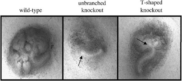
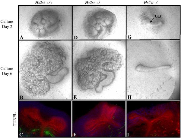
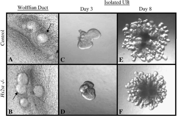
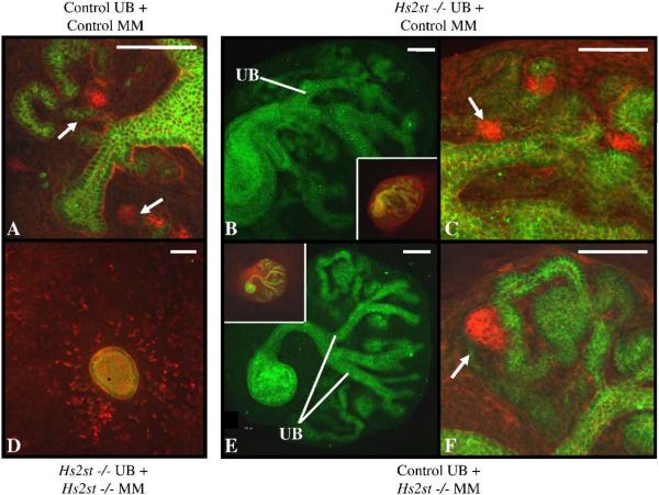
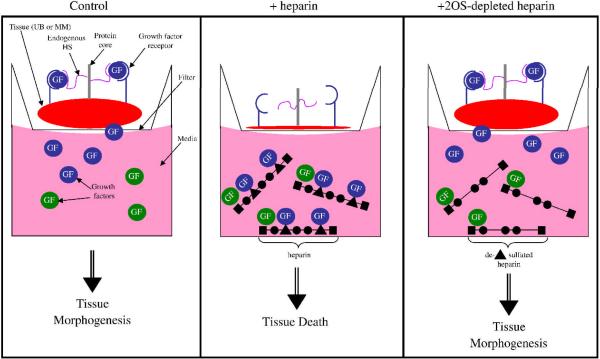
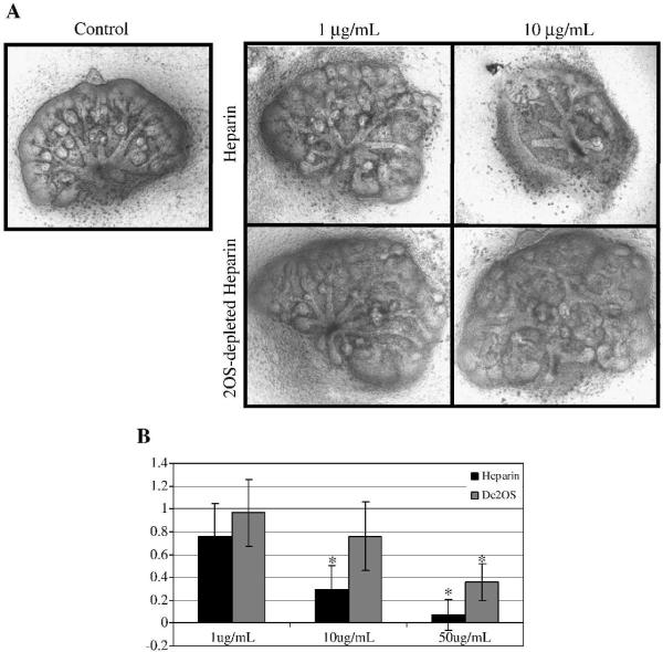
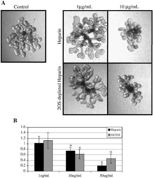
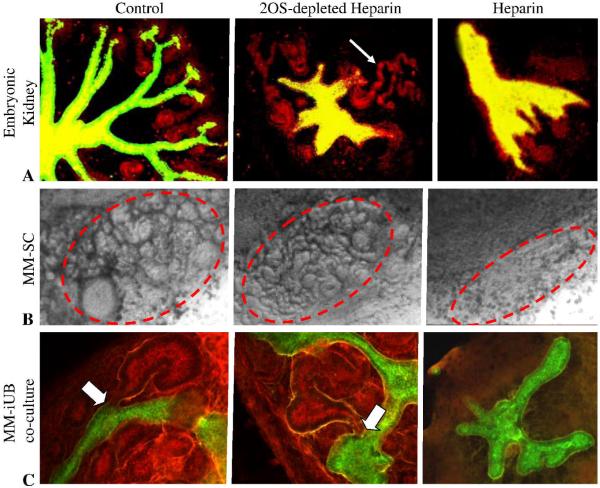
Heparan sulfate proteoglycans (HSPGs) are central modulators of developmental processes likely through their interaction with growth factors, such as GDNF, members of the FGF and TGFbeta superfamilies, EGF receptor ligands and HGF. Absence of the biosynthetic enzyme, heparan sulfate 2-O-sulfotransferase (Hs2st) leads to kidney agenesis. Using a novel combination of in vivo and in vitro approaches, we have reanalyzed the defect in morphogenesis of the Hs2st(-)(/)(-) kidney. Utilizing assays that separately model distinct stages of kidney branching morphogenesis, we found that the Hs2st(-/-) UB is able to undergo branching and induce mesenchymal-to-epithelial transformation when recombined with control MM, and the isolated Hs2st null UB is able to undergo branching morphogenesis in the presence of exogenous soluble pro-branching growth factors when embedded in an extracellular matrix, indicating that the UB is intrinsically competent. This is in contrast to the prevailing view that the defect underlying the renal agenesis phenotype is due to a primary role for 2-O sulfated HS in UB branching. Unexpectedly, the mutant MM was also fully capable of being induced in recombination experiments with wild-type tissue. Thus, both the mutant UB and mutant MM tissue appear competent in and of themselves, but the combination of mutant tissues fails in vivo and, as we show, in organ culture. We hypothesized a 2OS-dependent defect in the mutual inductive process, which could be on either the UB or MM side, since both progenitor tissues express Hs2st. In light of these observations, we specifically examined the role of the HS 2-O sulfation modification on the morphogenetic capacity of the UB and MM individually. We demonstrate that early UB branching morphogenesis is not primarily modulated by factors that depend on the HS 2-O sulfate modification; however, factors that contribute to MM induction are markedly sensitive to the 2-O sulfation modification. These data suggest that key defect in Hs2st null kidneys is the inability of MM to undergo induction either through a failure of mutual induction or a primary failure of MM morphogenesis. This results in normal UB formation but affects either T-shaped UB formation or iterative branching of the T-shaped UB (possibly two separate stages in collecting system development dependent upon HS). We discuss the possibility that a disruption in the interaction between HS and Wnts (e.g. Wnt 9b) may be an important aspect of the observed phenotype. This appears to be the first example of a defect in the MM preventing advancement of early UB branching past the first bifurcation stage, one of the limiting steps in early kidney development.


Figures

Figure 1
Figure 1
Phenotypic variations of Hs2st null kidneys. At e12.5, two different ureteric bud (UB) phenotypes are noted amongst Hs2st−/− embryonic kidneys. The most common phenotype is that of an unbranched UB (approximately two-thirds of kidneys analyzed) and the other is of a UB that has undergone the first dichotomous branching step (T-shaped, approximately one-third). Despite the fact that the UB has branched to a T-shape, overall branching is significantly delayed compared to wild-type and renal agenesis occurs at a 100% penetrance rate.
Figure 2
Figure 2
Embryonic kidney culture and TUNEL staining of kidney rudiments obtained from Hs2st heterozygous matings. Phase contrast micrographs of embryonic day 12 kidneys isolated and cultured in vitro for 6 days. Genotyping was performed following isolation. Both wild-type (A,B) and heterozygous (D,E) kidney rudiments display normal growth and branching patterns on culture day 2 and culture day 6. In contrast, knockout rudiments show no morphogenesis beyond outgrowth of the ureteric bud at culture day 2 (G; arrow points to the ureteric bud) and by culture day 6, the metanephric mesenchyme has degenerated although the ureter remains (H). All images at 10X. TUNEL staining in whole embryonic kidneys. Confocal micrographs of isolated wild-type (A), heterozygous (B) and knockout (C) embryonic day 11.5 kidneys stained for apoptotic cells (green), lotus lectin (red) and DAPI (blue). There is no significant difference in apoptosis between embryonic kidneys that express Hs2st (C,F) and embryonic kidneys that are deficient in Hs2st (H).
Figure 3
Figure 3
Hs2st expression in ureteric bud (UB) and metanephric mesenchyme (MM) is quantitatively similar at e11.5. Hs2st expression was analyzed using the GUDMAP database (www.gudmap.org). At e11.5, expression of Hs2st was quantitatively similar in the UB and MM while at e15.5, Hs2st expression is significantly higher in uninduced MM (peripheral blastema) vs. the UB tip. Average ± SD, n=3, *p<0.05 compared to e15.5 UB tip.
Figure 4
Figure 4
Epithelial morphogenesis occurs in Hs2st−/− Wolffian ducts (WD)and isolated ureteric buds (UB). A, B: Phase contrast micrographs of embryonic day 12 WD isolated and cultured in vitro for 3 days in DMEM/F12 supplemented with 125ng/ml GDNF and 125ng/ml FGF1. Genotyping was performed following isolation. Isolated WDs from control and knockout rudiments show similar levels of epithelial budding suggesting that the Hs2st null WD is competent to form a ureteric bud under these conditions. Arrows point to areas of budding from the WD. C–F: Phase contrast micrographs of UBs isolated from E11 kidney rudiments, suspended in a 3-dimensional extracellular matrix and cultured in the presence of BSN conditioned media and 125ng/ml GDNF and 125ng/ml FGF1. Isolated UBs from control (C,E) and knockout (D,F) rudiments show essentially the same pattern of growth and branching suggesting that the Hs2st null UB is competent to undergo branching morphogenesis under these conditions. All images at 10×.
Figure 5
Figure 5
Mix-and-match recombination culture between Hs2st knockout and control tissues. Ureteric buds (UB) and metanephric mesenchyme (MM) were isolated from E12 kidney rudiments. Knockout tissue was visually identified by lack of UB branching morphogenesis and tissue genotype was later confirmed via PCR. Confocal micrographs of recombined tissues stained with E-cadherin (green) and peanut agglutinin (red indicates podocytes) outline epithelial (green- UB and MM derived) and mesenchymal (red) structures. Images show lack of mutual induction in knockout UB recombined with knockout MM (D) and UB branching and mesenchymal-to-epithelial transformation in control (wild-type) UB recombined with control (wild-type) MM (A), knockout UB recombined with control (heterozygous) MM (B,C), and control (heterozygous) UB recombined with knockout MM, and (E,F). B,E: Epithelial structures in the recombined tissue, UB branches are noted emanating from the common ureter Insets show tissue with both green and red channels displayed. C,F: High power images of B,E showing pre-glomerular structures (arrows-podocytes) derived from the MM. Scale bars 100μm.
Figure 6
Figure 6
Schematic of the effect of heparin and 2OS-depleted heparin in the in vitro culture system. Under control conditions (far left panel), growth factors present within the media and secreted by the tissue of interest (embryonic kidney, ureteric bud or metanephric mesenchyme) are captured by heparan sulfate (HS) present on the cells and within the extracellular matrix of the tissue which results in tissue morphogenesis (blue circles represent growth factors necessary for morphogenesis while the green circles are not critical but may be supportive for morphogenesis). Middle panel: When fully sulfated heparin (with 2-O sulfated residues, represented by the triangle) is added to the media it binds a variety of growth factors. The heparin competitively binds factors away from endogenous HS, thereby trapping the factors within the media, resulting in tissue death. When 2OS-depleted heparin is added to the media (far left panel), growth factors that depend on the 2-O sulfated moiety for binding (blue circles), will be free in the media and able to bind to endogenous HS while those that do not depend on the 2-O sulfated moiety for binding (green circles) will continue to be trapped in the media. In this schematic, the blue growth factors are critical for tissue morphogenesis and are then able to induce morphogenesis.
Figure 7
Figure 7
A: Heparin and 2OS-depleted heparin titration curve on embryonic kidney rudiments. Phase contrast micrographs of e13 rat embryonic kidneys that were cultured in the presence of varying concentrations of heparin and 2OS-depleted heparin for 4–5 days. At a concentration of 10μg/mL, heparin markedly disrupts ureteric bud (UB) branching morphogenesis and mesenchymal induction while a similar dose of 2OS-depleted heparin has little effect on these processes. Images at 10× B: Graphical analysis of the average number of UB tips as a percentage of control. UB tips were counted after staining embryonic kidneys for UB specific Dolichos bifloris (raw data not shown). At higher concentrations (50 μg/mL), addition of heparin and 2OS-depleted heparin inhibits UB branching. Mean ± SD, n>3, *p<0.05 compared to control.
Figure 8
Figure 8
Heparin and 2OS-depleted heparin titration curve on isolated ureteric bud (UB) branching morphogenesis. A: Phase contrast micrographs of UBs isolated from E13 kidney rudiments, suspended in a 3-dimensional extracellular matrix and cultured in the presence of BSN conditioned media and 125ng/ml GDNF and 125ng/ml FGF1 and varying concentrations of heparin and 2OS-depleted heparin. Images at 10×. B: Graphical analysis of the average number of UB tips as a percentage of control. At a concentration of 10μg/mL and higher, both heparin and 2OS-depleted heparin significantly inhibit UB branching indicating that the binding of factors involved in UB branching are not markedly affected by the 2-O sulfate modification. Mean ± SD, n>3, *p<0.05 compared to control.
Figure 9
Figure 9
Effects of heparin and 2OS-depleted heparin on mesenchymal-to-epithelial transformation. A: A: Confocal micrographs of embryonic kidney cultured for 4 days in the absence (control) or presence of 100 μg/ml 2OS-depleted heparin or heparin. Note that addition of heparin results in generalized tissue death (A, far right panel) while addition of 2OS-depleted heparin (A, middle panel) inhibits UB branching but mesenchymal induction continues to take place at UB tips that do form (D. biflorus lectin (green) and E-cadherin (red); arrow point to induced MM structures) B: Phase contrast micrographs of metanephric mesenchyme (MM) isolated from E13 rat kidney co-cultured with spinal cord. MM was cultured for 5 days in the absence (control, far left panel) or presence of 100 mg/ml heparin (B, far right panel) or 2OS-depleted heparin (B, middle panel). Circled areas highlight MM that has undergone mesenchymal-to-epithelial transition. C: Effects of 2OS-depleted and fully sulfated heparin on mutual induction between the isolated UB and MM. Confocal micrographs of isolated UB cultured for 7 days then recombined with freshly isolated MM and further cultured for 5 days in the absence (control) or presence of 100 μg/ml heparin (C, far left panel) or 2OS-depleted heparin (C, middle panel) (D. biflorus (green) and peanut agglutinin (red) outline UB derived (green) and mesenchymal (red) structures). Note the absence of induced MM structures including comma and s-shaped bodies in the heparin treated tissues and apparently normal MM induction in the presence of 2OS-depleted heparin (arrowheads point to induced MM structures; arrows in higher magnification images (D) point to connecting segments between isolated UB and MM derived tissues).

Similar articles

Cited by

References

    1. Baeg GH, Selva EM, Goodman RM, Dasgupta R, Perrimon N. The Wingless morphogen gradient is established by the cooperative action of Frizzled and Heparan Sulfate Proteoglycan receptors. Dev Biol. 2004;276:89–100. - PubMed
    1. Brunskill EW, Aronow BJ, Georgas K, Rumballe B, Valerius MT, Aronow J, Kaimal V, Jegga AG, Yu J, Grimmond S, McMahon AP, Patterson LT, Little MH, Potter SS. Atlas of gene expression in the developing kidney at microanatomic resolution. Dev Cell. 2008;15:781–91. - PMC - PubMed
    1. Bullock SL, Fletcher JM, Beddington RS, Wilson VA. Renal agenesis in mice homozygous for a gene trap mutation in the gene encoding heparan sulfate 2-sulfotransferase. Genes Dev. 1998;12:1894–906. - PMC - PubMed
    1. Bush KT, Sakurai H, Steer DL, Leonard MO, Sampogna RV, Meyer TN, Schwesinger C, Qiao J, Nigam SK. TGF-beta superfamily members modulate growth, branching, shaping, and patterning of the ureteric bud. Dev Biol. 2004;266:285–98. - PubMed
    1. Cacalano G, Farinas I, Wang LC, Hagler K, Forgie A, Moore M, Armanini M, Phillips H, Ryan AM, Reichardt LF, Hynes M, Davies A, Rosenthal A. GFRalpha1 is an essential receptor component for GDNF in the developing nervous system and kidney. Neuron. 1998;21:53–62. - PMC - PubMed

Publication types

MeSH terms

Substances

LinkOut - more resources