Somatotopic organization in the internal segment of the globus pallidus in Parkinson's disease
- PMID: 20059997
- PMCID: PMC4920550
- DOI: 10.1016/j.expneurol.2009.12.030
Somatotopic organization in the internal segment of the globus pallidus in Parkinson's disease
Abstract
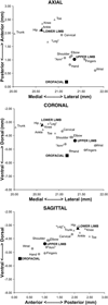
Ablation or deep brain stimulation in the internal segment of the globus pallidus (GPi) is an effective therapy for the treatment of Parkinson's disease (PD). Yet many patients receive only partial benefit, including varying levels of improvement across different body regions, which may relate to a differential effect of GPi surgery on the different body regions. Unfortunately, our understanding of the somatotopic organization of human GPi is based on a small number of studies with limited sample sizes, including several based upon only a single recording track or plane. To fully address the three-dimensional somatotopic organization of GPi, we examined the receptive field properties of pallidal neurons in a large cohort of patients undergoing stereotactic surgery. The response of neurons to active and passive movements of the limbs and orofacial structures was determined, using a minimum of three tracks across at least two medial-lateral planes. Neurons (3183) were evaluated from 299 patients, of which 1972 (62%) were modulated by sensorimotor manipulation. Of these, 1767 responded to a single, contralateral body region, with the remaining 205 responding to multiple and/or ipsilateral body regions. Leg-related neurons were found dorsal, medial and anterior to arm-related neurons, while arm-related neurons were dorsal and lateral to orofacial-related neurons. This study provides a more detailed map of individual body regions as well as specific joints within each region and provides a potential explanation for the differential effect of lesions or DBS of the GPi on different body parts in patients undergoing surgical treatment of movement disorders.
Copyright 2010 Elsevier Inc. All rights reserved.
Figures


References
-
- Alexander GE, DeLong MR, Strick PL. Parallel organization of functionally segregated circuits linking basal ganglia and cortex. Annu. Rev. Neurosci. 1986;9:357–381. - PubMed
-
- Asanuma C, Thach WR, Jones EG. Anatomical evidence for segregated focal groupings of efferent cells and their terminal ramifications in the cerebellothalamic pathway of the monkey. Brain Res. 1983;286:267–297. - PubMed
-
- Baron MS, Sidibe M, DeLong MR, Smith Y. Course of motor and associative pallidothalamic projections in monkeys. J. Comp. Neurol. 2001;429:490–501. - PubMed
-
- Bergman H, Wichmann T, DeLong MR. Reversal of experimental parkinsonism by lesions of the subthalamic nucleus. Science. 1990;249:1436–1438. - PubMed
MeSH terms
Grants and funding
LinkOut - more resources
Full Text Sources
Medical

