Comparative Study
. 2010 Apr 15;244(2):218-25.
doi: 10.1016/j.taap.2009.12.035. Epub 2010 Jan 6.

Transport of N-acetyl-S-(1,2-dichlorovinyl)-L-cysteine, a metabolite of trichloroethylene, by mouse multidrug resistance associated protein 2 (Mrp2)

Affiliations
Comparative Study

Transport of N-acetyl-S-(1,2-dichlorovinyl)-L-cysteine, a metabolite of trichloroethylene, by mouse multidrug resistance associated protein 2 (Mrp2)

Kirill Tsirulnikov et al. Toxicol Appl Pharmacol. .

Abstract

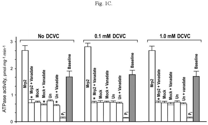
N-acetyl-S-(1,2-dichlorovinyl)-l-cysteine (Ac-DCVC) and S-(1,2-dichlorovinyl)-l-cysteine (DCVC) are the glutathione conjugation pathway metabolites of a common industrial contaminant and potent nephrotoxicant trichloroethylene (TCE). Ac-DCVC and DCVC are accumulated in the renal proximal tubule where they may be secreted into the urine by an unknown apical transporter(s). In this study, we explored the hypothesis that the apical transport of Ac-DCVC and/or DCVC may be mediated by the multidrug resistance associated protein 2 (Mrp2, ABCC2), which is known to mediate proximal tubular apical ATP-dependent transport of glutathione and numerous xenobiotics and endogenous substances conjugated with glutathione. Transport experiments using membrane vesicles prepared from mouse proximal tubule derived cells expressing mouse Mrp2 utilizing ATPase assay and direct measurements of Ac-DCVC/DCVC using liquid chromatography/tandem mass-spectrometry (LC/MS/MS) demonstrated that mouse Mrp2 mediates ATP-dependent transport of Ac-DCVC. Expression of mouse Mrp2 antisense mRNA significantly inhibited the vectorial basolateral to apical transport of Ac-DCVC but not DCVC in mouse proximal tubule derived cells endogenously expressing mouse Mrp2. The results suggest that Mrp2 may be involved in the renal secretion of Ac-DCVC.


Conflict of interest statement

Conflict of interests statement: The authors have no conflict of interests to state.

Figures

Fig. 1
Fig. 1
(A) Quantification of mouse Mrp2 in the membrane vesicles prepared from the mPCT-A2 cells (mPCT-A2) transiently expressing mouse Mrp2 (Ex) and from control untransfected (Un) and mock-transfected (Mock) mPCT-A2 cells were resolved on 7.5% SDS-PAGE, transferred onto PDF membranes and probed with RP-4 mouse Mrp2 specific antibody (IgG) and with the same antibody preincubated with the peptide (10:1) used for immunization (IgG + P). Loading: 7 μg. mPCT cells (mPCT) endogenously express Mrp2. The membranes were isolated from mPCT cells using the technique described for mPCT-A2 cell membrane isolation, resolved on 7.5% SDS-PAGE and immunoblotted with RP-4 antibody (IgG) and with the same antibody preincubated with the peptide used for immunization (IgG + P). Loading: 50 μg. The effect of Ac-DCVC (B) and DCVC (C) on ATPase activity of the membrane vesicles prepared from mPCT-A2 cells expressing mouse Mrp2 (Mrp2), mock-transfected (Mock) or untransfected (Un) mPCT-A2 cells without and in the presence of 1.2 mM vanadate (Vanadate). ATPase activity was measured in an assay containing 5 mM ATP, 10 mM MgCl2, 50 mM KCl, 2 mM GSH, 2 mM dithiothreitol, and 50 mM HEPES, pH 7.5. The amount of inorganic phosphate formed nonenzymatically in the assay was determined in the same assay containing 2% SDS (Pi). The 0.1 mM or 1 mM compound was added into the ATPase assay without preincubation of vesicles. * Shows significant difference (p<0.05) of the total ATPase activity of the vesicles prepared from the cells expressing Mrp2 (or mock-transfected or untransfected cells) without vanadate to the same activity in the presence of 1.2 mM vanadate. (D) Effect of estradiol 17-β-D-glucuronide (E17bG), leukotriene C4 (LTC4), pronencid, Ac-DCVC, DCVC, N-acetyl-S-(2,2-dichlorovinyl)-L-cysteine (Ac-2,2-DCVC), N-acetyl-S-(4-chlorobenzyl)-L-cysteine (Ac-4-CBC), N-acetyl-S-(4-bromobenzyl)-L-cysteine (Ac-4-BBC), N-acetyl-S-(1,1-difluoro-2,2-dichloroethyl)-L-cysteine (Ac-1,1-DF-2,2-DCC), N-acetyl-S-(1,1-dichloro-2,2-difluoroethyl)-L-cysteine (Ac-1,1-DC-2,2-DFC), N-acetyl-L-cysteine (Ac-Cys) and N-acetyl-L-tyrosine (Ac-Tyr) of the baseline ATPase activity (None) of membrane vesicles prepared from the mPCT-A2 cells expressing Mrp2. The 0.1 mM or 1 mM compound (black and white bars, respectively) was added into the ATPase assay. LTC4 was used at a concentration of 4 μM. The ATPase activity, measured in the presence of compound, is shown as a ratio to the baseline activity without compound (None). * Significant difference (p<0.05) from the baseline activity. (E) Inhibition of baseline ATPase activity of membrane vesicles prepared from untransfected and Mrp2 expressing mPCT-A2 cells by anti-Mrp2 antibody. Vesicles (100 μg) were pretreated at 37°C for 30 min with RP-4 antibody (100 μg) before ATPase activity was measured as described in Materials and methods. * Shows significant difference (p<0.05) between ATPase activity of untreated membrane vesicles in comparison with the vesicles incubated with RP-4 antibody.
Fig. 1
Fig. 1
(A) Quantification of mouse Mrp2 in the membrane vesicles prepared from the mPCT-A2 cells (mPCT-A2) transiently expressing mouse Mrp2 (Ex) and from control untransfected (Un) and mock-transfected (Mock) mPCT-A2 cells were resolved on 7.5% SDS-PAGE, transferred onto PDF membranes and probed with RP-4 mouse Mrp2 specific antibody (IgG) and with the same antibody preincubated with the peptide (10:1) used for immunization (IgG + P). Loading: 7 μg. mPCT cells (mPCT) endogenously express Mrp2. The membranes were isolated from mPCT cells using the technique described for mPCT-A2 cell membrane isolation, resolved on 7.5% SDS-PAGE and immunoblotted with RP-4 antibody (IgG) and with the same antibody preincubated with the peptide used for immunization (IgG + P). Loading: 50 μg. The effect of Ac-DCVC (B) and DCVC (C) on ATPase activity of the membrane vesicles prepared from mPCT-A2 cells expressing mouse Mrp2 (Mrp2), mock-transfected (Mock) or untransfected (Un) mPCT-A2 cells without and in the presence of 1.2 mM vanadate (Vanadate). ATPase activity was measured in an assay containing 5 mM ATP, 10 mM MgCl2, 50 mM KCl, 2 mM GSH, 2 mM dithiothreitol, and 50 mM HEPES, pH 7.5. The amount of inorganic phosphate formed nonenzymatically in the assay was determined in the same assay containing 2% SDS (Pi). The 0.1 mM or 1 mM compound was added into the ATPase assay without preincubation of vesicles. * Shows significant difference (p<0.05) of the total ATPase activity of the vesicles prepared from the cells expressing Mrp2 (or mock-transfected or untransfected cells) without vanadate to the same activity in the presence of 1.2 mM vanadate. (D) Effect of estradiol 17-β-D-glucuronide (E17bG), leukotriene C4 (LTC4), pronencid, Ac-DCVC, DCVC, N-acetyl-S-(2,2-dichlorovinyl)-L-cysteine (Ac-2,2-DCVC), N-acetyl-S-(4-chlorobenzyl)-L-cysteine (Ac-4-CBC), N-acetyl-S-(4-bromobenzyl)-L-cysteine (Ac-4-BBC), N-acetyl-S-(1,1-difluoro-2,2-dichloroethyl)-L-cysteine (Ac-1,1-DF-2,2-DCC), N-acetyl-S-(1,1-dichloro-2,2-difluoroethyl)-L-cysteine (Ac-1,1-DC-2,2-DFC), N-acetyl-L-cysteine (Ac-Cys) and N-acetyl-L-tyrosine (Ac-Tyr) of the baseline ATPase activity (None) of membrane vesicles prepared from the mPCT-A2 cells expressing Mrp2. The 0.1 mM or 1 mM compound (black and white bars, respectively) was added into the ATPase assay. LTC4 was used at a concentration of 4 μM. The ATPase activity, measured in the presence of compound, is shown as a ratio to the baseline activity without compound (None). * Significant difference (p<0.05) from the baseline activity. (E) Inhibition of baseline ATPase activity of membrane vesicles prepared from untransfected and Mrp2 expressing mPCT-A2 cells by anti-Mrp2 antibody. Vesicles (100 μg) were pretreated at 37°C for 30 min with RP-4 antibody (100 μg) before ATPase activity was measured as described in Materials and methods. * Shows significant difference (p<0.05) between ATPase activity of untreated membrane vesicles in comparison with the vesicles incubated with RP-4 antibody.
Fig. 2
Fig. 2
(A) The ATP-dependent transport of Ac-DCVC (black bars) and DCVC (grey bars) into membrane vesicles prepared from the mPCT cells expressing mouse Mrp2 (Ex) measured by LC/MS/MS. ATP was replaced with an equimolar concentration of AMP in control experiments (white bars). * Significant difference (p<0.05) from control with AMP instead of ATP. (B) A representative curve of ATP-dependent Ac-DCVC transport inside Mrp2 vesicles. The data are means ± S.D. of two measurements. (C) The Lineweaver-Burk plot of the data shown in (B). S0.5 values were determined as the substrate concentration at half-maximal velocity of transport under the experimental conditions described above using the double-reciprocal plot (Lineweaver, Burk, 1934).
Fig. 2
Fig. 2
(A) The ATP-dependent transport of Ac-DCVC (black bars) and DCVC (grey bars) into membrane vesicles prepared from the mPCT cells expressing mouse Mrp2 (Ex) measured by LC/MS/MS. ATP was replaced with an equimolar concentration of AMP in control experiments (white bars). * Significant difference (p<0.05) from control with AMP instead of ATP. (B) A representative curve of ATP-dependent Ac-DCVC transport inside Mrp2 vesicles. The data are means ± S.D. of two measurements. (C) The Lineweaver-Burk plot of the data shown in (B). S0.5 values were determined as the substrate concentration at half-maximal velocity of transport under the experimental conditions described above using the double-reciprocal plot (Lineweaver, Burk, 1934).
Fig. 3
Fig. 3
(A) Apical to basolateral and (B) basolateral to apical transport of Ac-DCVC through the monolayer of mPCT cells (white bars) or the mPCT cells transfected with anti-mouse Mrp2 shRNA plasmid (black bars). In control experiments (C) mPCT cells were transfected with a nonspecific anti-AA3 shRNA plasmid (Fig. 3C). Note that mPCT cells do not express AA3 (Newman et al., 2007). (D) Anti-Mrp2 shRNA significantly decreased the expression of Mrp2 protein in mPCT cells in comparison with untransfected cells (control) whereas anti-AA3 shRNA did not. A representative immunoblot is shown on the left. The membranes were isolated from untransfected (Control) and transfected with AA3 and Mrp2 shRNA plasmids mPCT cells using the technique described for mPCT-A2 cell membrane isolation, resolved on 7.5% SDS-PAGE and immunoblotted with RP-4 antibody. Loading: 50 μg. Graphic illustration of Mrp2 quantification in 3 separate experiments is shown on the right. The bands were scanned and quantified in Adobe Photoshop (Adobe Instruments). * Shows significant difference (p<0.05) between transfected and untransfected (control) cells. (E) Apical to basolateral and basolateral to apical transport of DCVC through the monolayer of mPCT cells (white bars) and the mPCT cells transfected with anti-mouse Mrp2 shRNA plasmid (black bars). * Shows significant difference (p<0.05) between transfected and untransfected (control) cells.
Fig. 3
Fig. 3
(A) Apical to basolateral and (B) basolateral to apical transport of Ac-DCVC through the monolayer of mPCT cells (white bars) or the mPCT cells transfected with anti-mouse Mrp2 shRNA plasmid (black bars). In control experiments (C) mPCT cells were transfected with a nonspecific anti-AA3 shRNA plasmid (Fig. 3C). Note that mPCT cells do not express AA3 (Newman et al., 2007). (D) Anti-Mrp2 shRNA significantly decreased the expression of Mrp2 protein in mPCT cells in comparison with untransfected cells (control) whereas anti-AA3 shRNA did not. A representative immunoblot is shown on the left. The membranes were isolated from untransfected (Control) and transfected with AA3 and Mrp2 shRNA plasmids mPCT cells using the technique described for mPCT-A2 cell membrane isolation, resolved on 7.5% SDS-PAGE and immunoblotted with RP-4 antibody. Loading: 50 μg. Graphic illustration of Mrp2 quantification in 3 separate experiments is shown on the right. The bands were scanned and quantified in Adobe Photoshop (Adobe Instruments). * Shows significant difference (p<0.05) between transfected and untransfected (control) cells. (E) Apical to basolateral and basolateral to apical transport of DCVC through the monolayer of mPCT cells (white bars) and the mPCT cells transfected with anti-mouse Mrp2 shRNA plasmid (black bars). * Shows significant difference (p<0.05) between transfected and untransfected (control) cells.

Similar articles

Cited by

References

    1. Agency for Toxic Substances and Disease Registry (ATSDR) Toxicological Profile for Trichloroethylene. U.S. Public Health Service, U.S. Department of Health and Human Services; Atlanta, GA: 1993. - PubMed
    1. Anders MW, Dekant W. Glutathione-dependent bioactivation of haloalkenes. Annu Rev Pharmacol Toxicol. 1998;38:501–537. - PubMed
    1. Anders MW, Dekant W, Vamvakas S. Formation and fate of nephrotoxic and cytotoxic glutathione S-conjugates: cysteine conjugate beta-lyase pathway. Adv Pharmacol. 1994;27:115–162. - PubMed
    1. Bencini DA, Wild JR, O'Donovan GA. Linear one-step assay for the determination of orthophosphate. Anal Biochem. 1983;132:254–258. - PubMed
    1. Birner G, Bernauer U, Werner M, Dekant W. Biotransformation, excretion, and nephrotoxicity of haloalkene-derived cysteine S-conjugates. Arch Toxicol. 1997;72:1–8. - PubMed

Publication types

MeSH terms

Substances