

Development and optimization of a doxorubicin loaded poly(lactic acid) contrast agent for ultrasound directed drug delivery
- PMID: 20060024
- PMCID: PMC2878717
- DOI: 10.1016/j.jconrel.2009.12.021
Development and optimization of a doxorubicin loaded poly(lactic acid) contrast agent for ultrasound directed drug delivery
Abstract
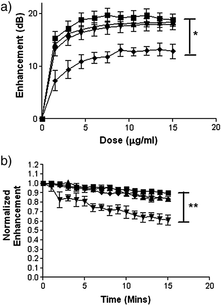
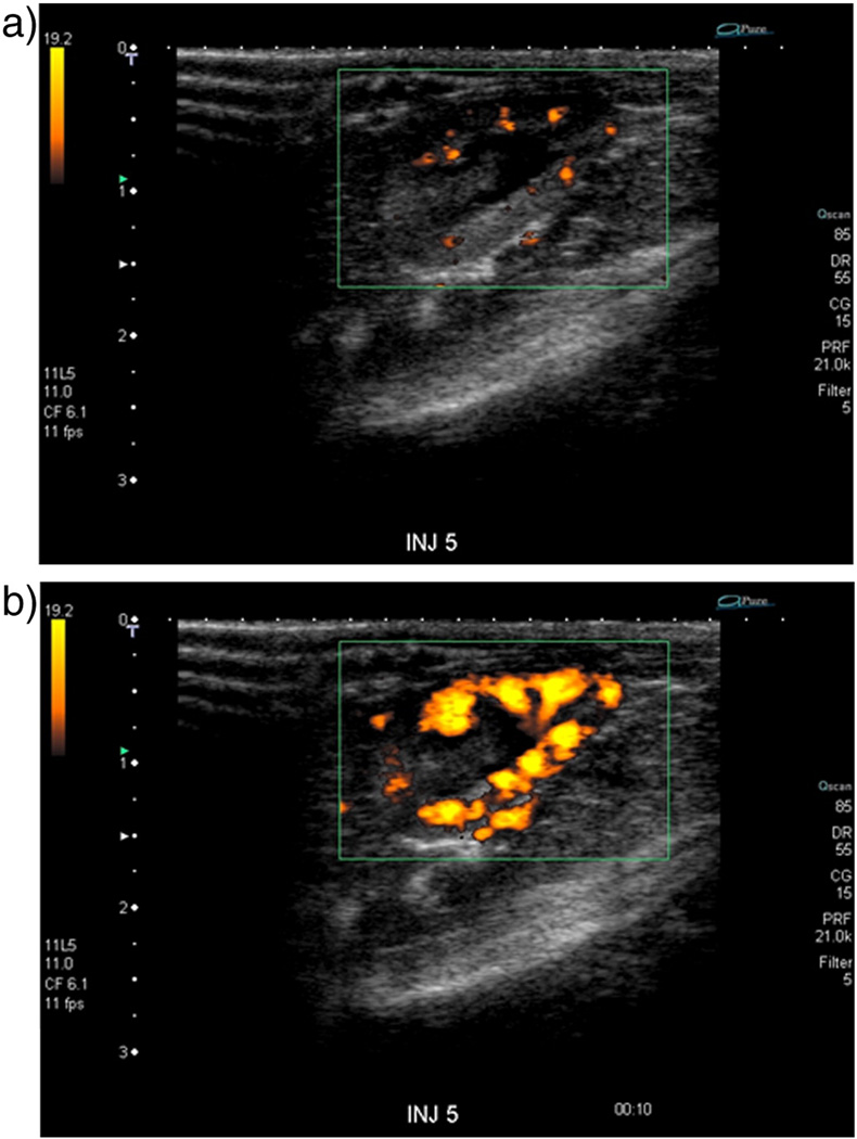
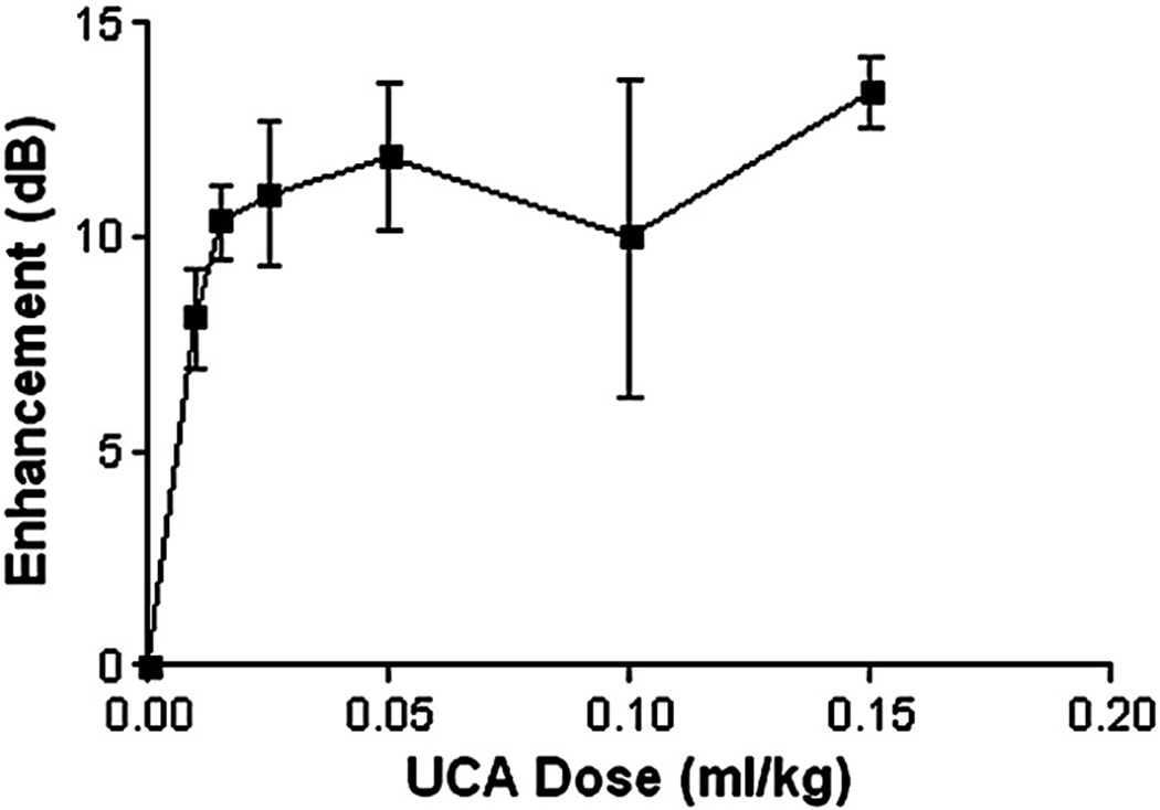
An echogenic, intravenous drug delivery platform is proposed in which an encapsulated chemotherapeutic can travel to a desired location and drug delivery can be triggered using external, focused ultrasound at the area of interest. Three methods of loading poly(lactic acid) (PLA) shelled ultrasound contrast agents (UCA) with doxorubicin are presented. Effects on encapsulation efficiency, in vitro enhancement, stability, particle size, morphology and release during UCA rupture are compared by loading method and drug concentration. An agent containing doxorubicin within the shell was selected as an ideal candidate for future hepatocellular carcinoma studies. The agent achieved a maximal drug load of 6.2 mg Dox/g PLA with an encapsulation efficiency of 20.5%, showed a smooth surface morphology and tight size distribution (poly dispersity index=0.309) with a peak size of 1865 nm. Acoustically, the agent provided 19 dB of enhancement in vitro at a dosage of 10 microg/ml, with a half life of over 15 min. In vivo, the agent provided ultrasound enhancement of 13.4+/-1.6 dB within the ascending aorta of New Zealand rabbits at a dose of 0.15 ml/kg. While the drug-incorporated agent is thought to be well suited for future drug delivery experiments, this study has shown that agent properties can be tailored for specific applications based on choice of drug loading method.
Copyright 2009 Elsevier B.V. All rights reserved.
Figures
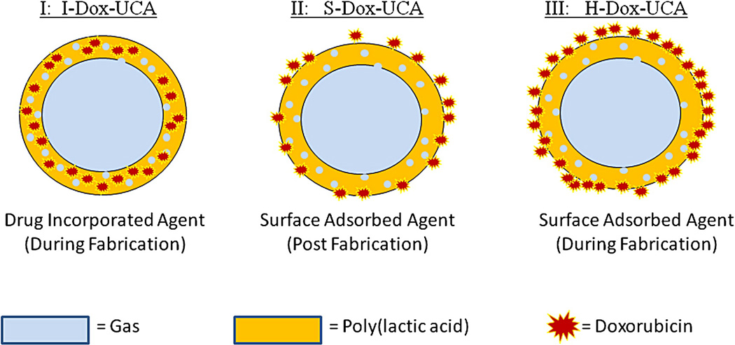
 , I-Dox-UCA =
  , S-Dox-UCA =
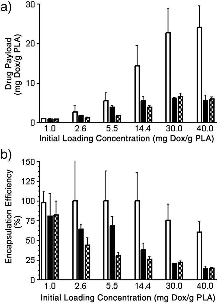
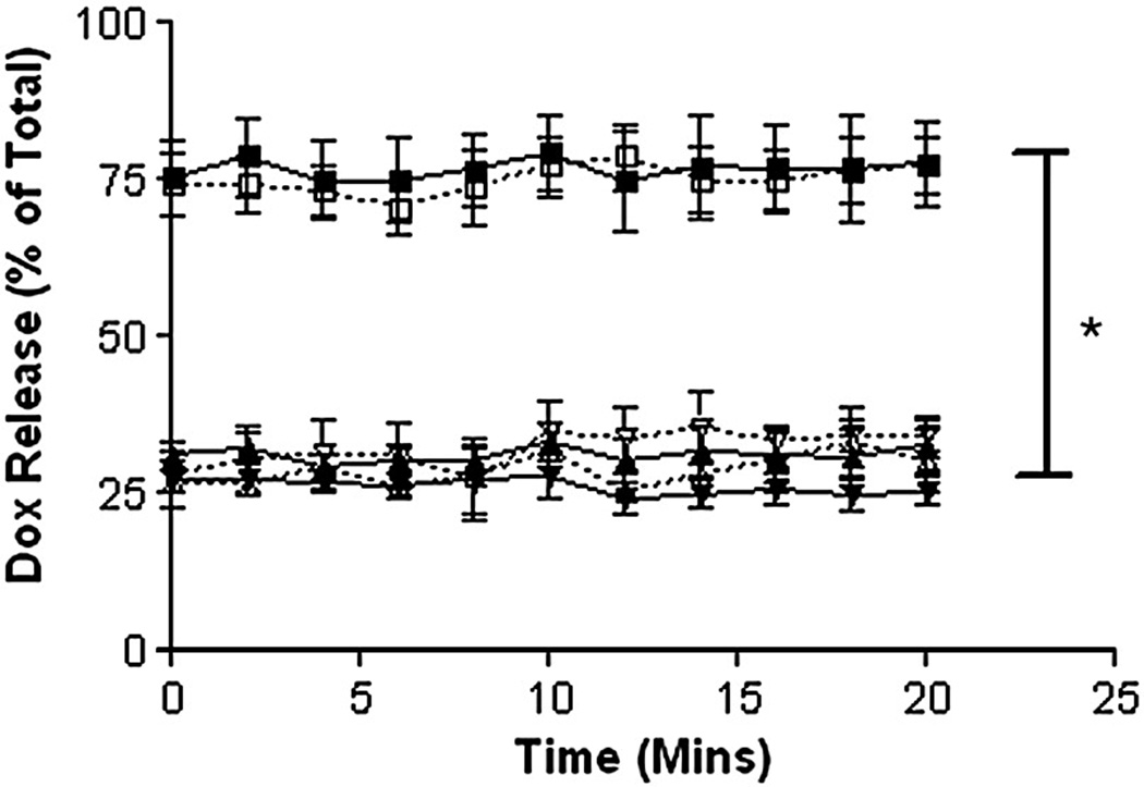
  . H-Dox-UCA approached a maximal drug load of 24.1 mg Dox/g PLA (encapsulation efficiency of 60.2%) at an initial loading concentration of 40.0 mg Dox/g PLA. Both the I-Dox-UCA and S-Dox-UCA samples reached peak drug payloads of 6.2 and 6.5 mg Dox/g PLA (encapsulation efficiencies of 20.5 and 21.9%) respectively at an initial loading concentration of 30.0 mg Dox/g PLA.
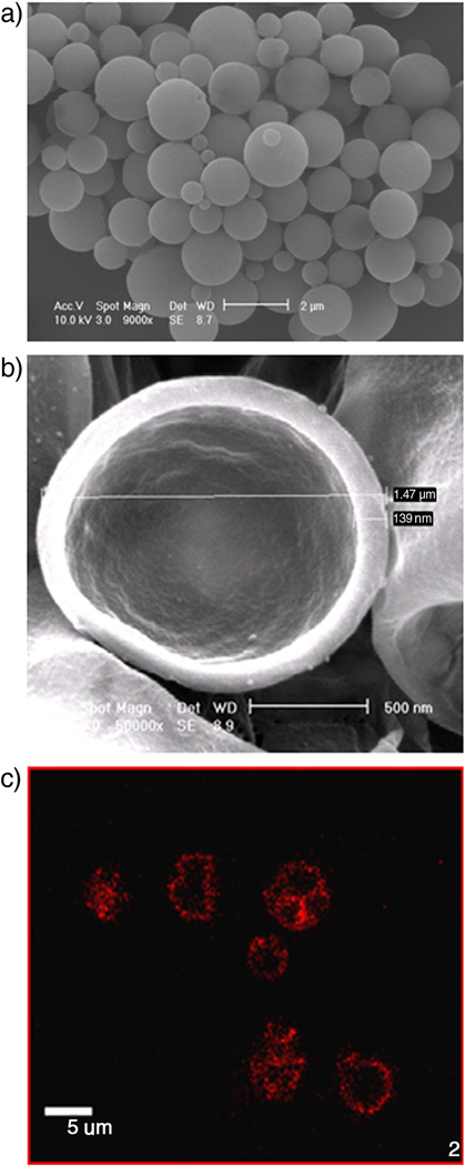
Similar articles
-
Ultrasound triggered cell death in vitro with doxorubicin loaded poly lactic-acid contrast agents.Ultrasonics. 2009 Dec;49(8):628-33. doi: 10.1016/j.ultras.2009.03.003. Epub 2009 Mar 28. Ultrasonics. 2009. PMID: 19394992 Free PMC article.
-
Delivery of encapsulated Doxorubicin by ultrasound-mediated size reduction of drug-loaded polymer contrast agents.IEEE Trans Biomed Eng. 2010 Jan;57(1):24-8. doi: 10.1109/TBME.2009.2030497. Epub 2009 Aug 25. IEEE Trans Biomed Eng. 2010. PMID: 19709952
-
Plasma sterilization of poly lactic acid ultrasound contrast agents: surface modification and implications for drug delivery.Ultrasound Med Biol. 2009 Nov;35(11):1854-62. doi: 10.1016/j.ultrasmedbio.2009.06.1098. Epub 2009 Sep 19. Ultrasound Med Biol. 2009. PMID: 19766380 Free PMC article.
-
New doxorubicin-loaded phospholipid microbubbles for targeted tumor therapy: Part I--Formulation development and in-vitro characterization.J Control Release. 2010 Apr 2;143(1):143-50. doi: 10.1016/j.jconrel.2009.12.026. Epub 2010 Jan 11. J Control Release. 2010. PMID: 20060861
-
Preparation, characterization and in vitro cytotoxicity of indomethacin-loaded PLLA/PLGA microparticles using supercritical CO2 technique.Eur J Pharm Biopharm. 2008 Sep;70(1):85-97. doi: 10.1016/j.ejpb.2008.03.011. Epub 2008 Mar 29. Eur J Pharm Biopharm. 2008. PMID: 18495445
Cited by
-
Polymeric Microbubble Shell Engineering: Microporosity as a Key Factor to Enhance Ultrasound Imaging and Drug Delivery Performance.Adv Sci (Weinh). 2024 Oct;11(40):e2404385. doi: 10.1002/advs.202404385. Epub 2024 Aug 29. Adv Sci (Weinh). 2024. PMID: 39207095 Free PMC article.
-
Acoustic Parameters for Optimal Ultrasound-Triggered Release from Novel Spinal Hardware Devices.Ultrasound Med Biol. 2020 Feb;46(2):350-358. doi: 10.1016/j.ultrasmedbio.2019.10.002. Epub 2019 Nov 13. Ultrasound Med Biol. 2020. PMID: 31732196 Free PMC article.
-
State-of-the-art materials for ultrasound-triggered drug delivery.Adv Drug Deliv Rev. 2014 Jun;72:3-14. doi: 10.1016/j.addr.2013.12.010. Epub 2013 Dec 31. Adv Drug Deliv Rev. 2014. PMID: 24389162 Free PMC article. Review.
-
Ultrasound imaging beyond the vasculature with new generation contrast agents.Wiley Interdiscip Rev Nanomed Nanobiotechnol. 2015 Jul-Aug;7(4):593-608. doi: 10.1002/wnan.1326. Epub 2015 Jan 8. Wiley Interdiscip Rev Nanomed Nanobiotechnol. 2015. PMID: 25580914 Free PMC article. Review.
-
Ultrasound-responsive polymer-based drug delivery systems.Drug Deliv Transl Res. 2021 Aug;11(4):1323-1339. doi: 10.1007/s13346-021-00963-0. Epub 2021 Mar 24. Drug Deliv Transl Res. 2021. PMID: 33761101 Free PMC article. Review.
References
-
- Singal P, Iliskovic N. Doxorubicin-induced cardiomyopathy. New Engl. J. Med. 1998;339:900–905. - PubMed
-
- Alberts DS, Garcia DJ. Safety aspects of pegylated liposomal doxorubicin in patients with cancer. Drugs. 1997;54:30–35. - PubMed
-
- Goram AL, Richmond PL. Pegylated liposomal doxorubicin: tolerability and toxicity. Pharmacotherapy. 2001;21:751–763. - PubMed
-
- Hoff L. Acoustic properties of ultrasonic contrast agents. Ultrasonics. 1996;34:591–593.
-
- Goldberg BB, Raichlen JS, Forsberg F. Ultrasound contrast agents: basic principles and clinical applications. second edition. London: Martin Dunitz; 2001.
Publication types
MeSH terms
Substances
Grants and funding
LinkOut - more resources
Full Text Sources
Other Literature Sources
Miscellaneous
