

doi: 10.1016/j.ymeth.2009.12.010.
Epub 2010 Jan 6.
Examining the contents of isolated Xenopus germinal vesicles
Affiliations
- PMID: 20060047
- PMCID: PMC2868088
- DOI: 10.1016/j.ymeth.2009.12.010
Item in Clipboard
Examining the contents of isolated Xenopus germinal vesicles
Methods.
2010 May.
Abstract
One can manually isolate the giant oocyte nucleus or germinal vesicle (GV) of Xenopus from a living oocyte with nothing more complicated than jewelers' forceps and a dissecting microscope. Similarly, one can remove the nuclear envelope by hand and allow the lampbrush chromosomes and other nuclear organelles to spread on a microscope slide. After centrifugation, the nuclear contents adhere tightly to the slide, where they can be subjected to immunostaining or fluorescent in situ hybridization for visualization by conventional or confocal microscopy. Preparations of isolated GV contents reveal details of nuclear structure that are almost impossible to attain by more conventional techniques.
Figures
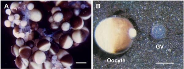
“Living” oocytes and GV of X. laevis. A. A small portion of an ovary from an adult female, in OR2 medium. Oocytes in various stages of development are present. In the largest oocytes the animal hemisphere is black or brown due to pigment that covers the yolk, whereas the white or yellowish-white yolk is visible in the vegetal hemisphere. Bar = I mm. B. A single oocyte (left) and its nearly transparent GV (right), which was squeezed out through a small hole in the animal pole. Some yolk extrudes through the hole. This GV was isolated in oil, but its appearance is similar to a GV isolated in a saline solution. Reproduced with permission from Paine et al. [27]. Bar = 500 μm.
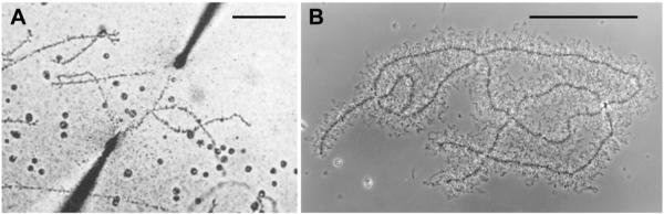
Giant LBCs from salamanders, isolated in a saline solution. A. The earliest image of a LBC isolated in a saline solution, from the newt Cynops (Triturus) pyrrhogaster. The homologous chromosomes are being stretched between microneedles. Reproduced from Duryee [6] with permission from the University of Pennsylvania Press. B. A pair of homologous LBCs from the newt Notophthalmus viridescens, isolated in a saline solution but not centrifuged. This image was taken by flash photography to stop the vigorous Brownian motion of the chromatin loops that extend laterally from the chromosome axis. Bars = 100 μm.
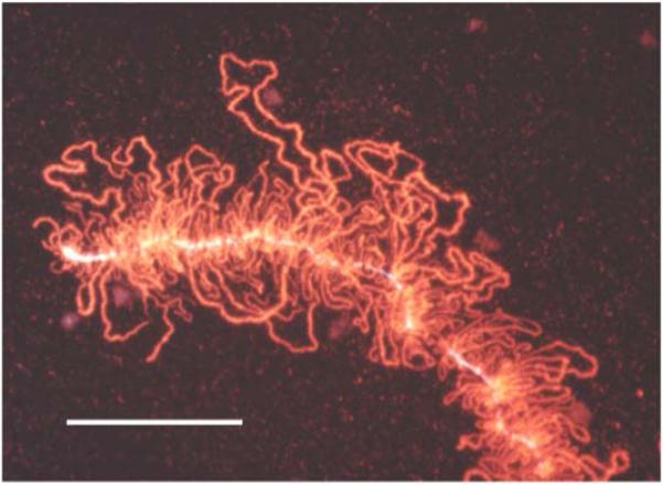
Effect of centrifugation on a LBC. A small segment of a LBC of the newt N. viridescens, from a preparation that had been centrifuged to attach the chromosomes to a glass microscope slide. Note that all the lateral loops of the chromosome are in essentially the same plane. This image was taken after immunofluorescent staining with an antibody against an abundant chromosomal protein. Bar = 50 μm.
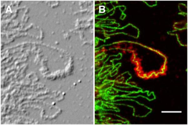
LBC preparations reveal details of chromosome structure at the molecular level. A. Differential interference contrast image of LBCs from the newt N. viridescens. B. The same region showing immunostaining with an antibody against RNA polymerase II (green) and cleavage stimulation factor 77 (red). Note that the green polymerase stain is limited to a diffraction-limited line in each lateral loop, especially well shown in the large loop in the center of the image. This line delineates the actively transcribing RNA polymerase molecules associated with the DNA axis of the loop. The red stain shows the nascent transcripts that coat the DNA axis. Modified from [32] with permission from Molecular Biology of the Cell. Bar = 5 μm.
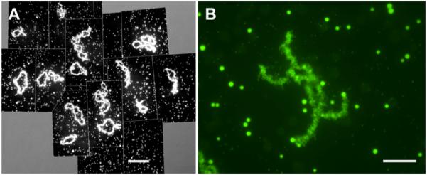
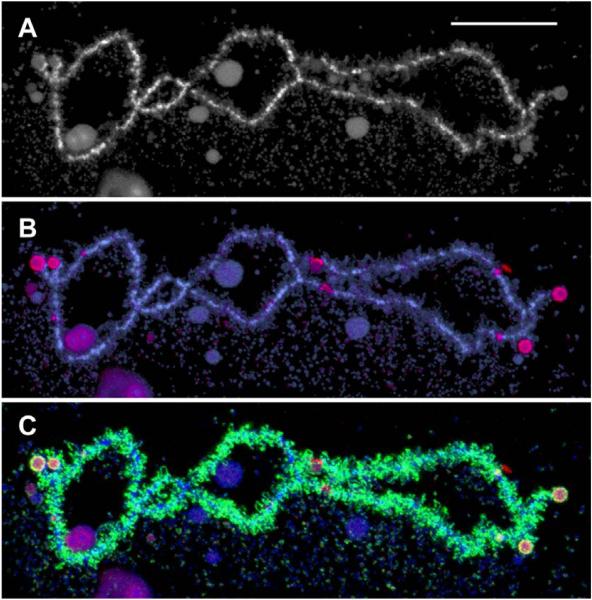
GV contents of X. laevis. A. Overall image of the contents of a single GV, showing the 18 lampbrush bivalents and several thousand nuclear bodies - primarily amplified nucleoli, Cajal bodies, and interchromatin granule clusters (speckles). Stained with mAb Y12, which reacts with small nuclear ribonucleoproteins. Bar = 100 μm. B. A single lampbrush bivalent with many nuclear speckles in the same field. Immunostained with mAb H5, which detects polymerase II in the chromosome loops and an unidentified epitope in the speckles. Bar = 10 μm.
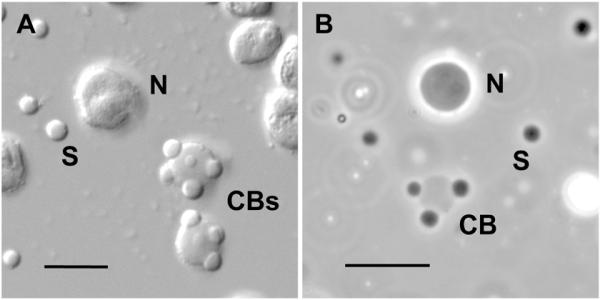
High magnification image of amplified nucleoli, Cajal bodies, and speckles. A. Differential interference contrast of nuclear organelles centrifuged onto a microscope slide after isolation in a saline solution. Each GV contains about 1000 nucleoli (N), 50-100 Cajal bodies (CBs) and several thousand interchromatin granule clusters or speckles (S). The contrast is high because the bodies are viewed in a medium of low refractive index. B. Phase contrast image of unfixed nuclear organelles in an oil-isolated GV. The organelles appear of low optical contrast because they are still in the nucleoplasm, which has a relatively high refractive index. As shown by fluorescence recovery after photobleaching (FRAP) studies, these organelles still exhibit active exchange of macromolecules with the surrounding medium [33, 34]. Panel B reproduced from [29] with permission from Molecular Biology of the Cell. Bars = 10 μm.
A single LBC from a mature GV of X. tropicalis. An advantage of X. tropicalis over X. laevis for the study of LBCs is the smaller chromosome number (n = 10 vs n = 18) and the fact that the genome has been sequenced (http://www.xenbase.org ). Working maps of the LBCs have also been published recently [35]. A. The longest chromosome of the set, stained with the DNA-specific dye DAPI. B. Immunofluorescent staining (red) with an antibody that recognizes RNA polymerase III. A few interstitial sites of pol III transcription are evident. The nature of the stained spherical objects on the chromosome ends is not known. Counterstained with DAPI (blue). C. The same chromosome showing staining for pol II (green) and pol III (red). The majority of loops are transcribed by pol II. Bar = 20 μm.
References
-
- Purkinje JE. Symbolae ad ovi avium historiam ante incubationem. Leopold Vossi; Leipzig: 1830.
-
- Brown R. Trans. Linnean Soc. Lond. 1833;16:685–745.
-
- Flemming W. Zellsubstanz, Kern und Zelltheilung. F. C. W. Vogel; Leipzig: 1882.
-
- Rückert J. Anat. Anz. 1892;7:107–158.
-
- Duryee WR. Arch. exp. Zellforsch. 1937;19:171–176.
Publication types
MeSH terms
Grants and funding
LinkOut - more resources
Full Text Sources
