. 2010 Jan 29;32(1):54-66.
doi: 10.1016/j.immuni.2009.12.003. Epub 2010 Jan 7.

The receptor SIGIRR suppresses Th17 cell proliferation via inhibition of the interleukin-1 receptor pathway and mTOR kinase activation

Affiliations

The receptor SIGIRR suppresses Th17 cell proliferation via inhibition of the interleukin-1 receptor pathway and mTOR kinase activation

Muhammet F Gulen et al. Immunity. .

Abstract

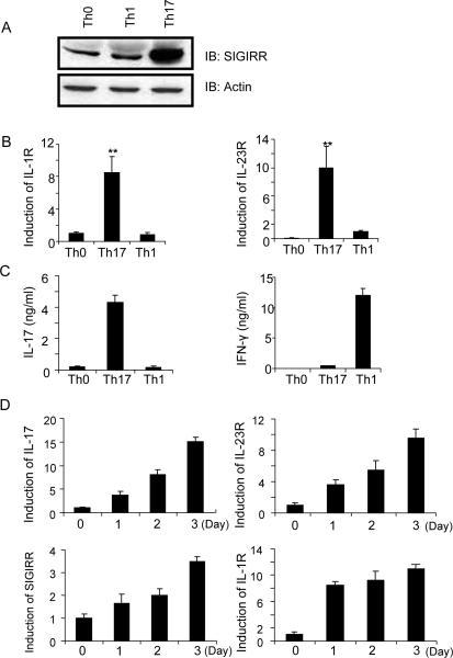
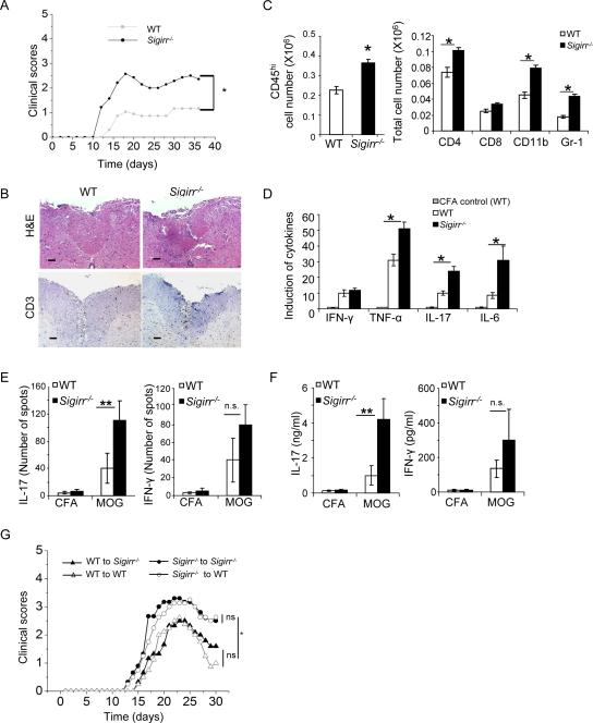
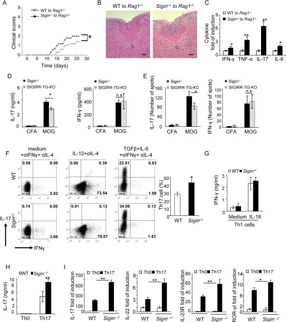
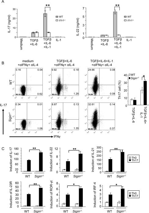
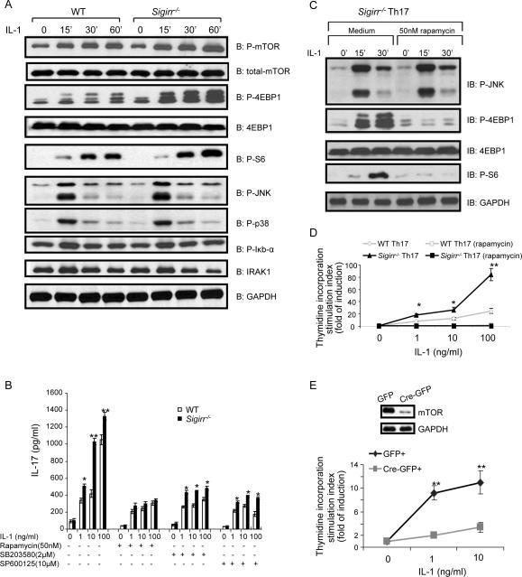
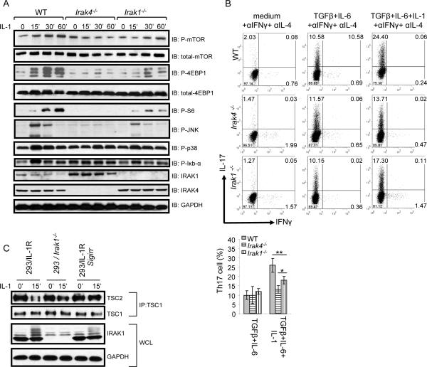
Interleukin-1 (IL-1)-mediated signaling in T cells is essential for T helper 17 (Th17) cell differentiation. We showed here that SIGIRR, a negative regulator of IL-1 receptor and Toll-like receptor signaling, was induced during Th17 cell lineage commitment and governed Th17 cell differentiation and expansion through its inhibitory effects on IL-1 signaling. The absence of SIGIRR in T cells resulted in increased Th17 cell polarization in vivo upon myelin oligodendrocyte glycoprotein (MOG(35-55)) peptide immunization. Recombinant IL-1 promoted a marked increase in the proliferation of SIGIRR-deficient T cells under an in vitro Th17 cell-polarization condition. Importantly, we detected increased IL-1-induced phosphorylation of JNK and mTOR kinase in SIGIRR-deficient Th17 cells compared to wild-type Th17 cells. IL-1-induced proliferation was abolished in mTOR-deficient Th17 cells, indicating the essential role of mTOR activation. Our results demonstrate an important mechanism by which SIGIRR controls Th17 cell expansion and effector function through the IL-1-induced mTOR signaling pathway.


Figures

Figure 1
Figure 1. SIGIRR expression is induced during Th17 cell differentiation
(A–C) Naïve CD4+ T cells (CD4+CD44lo) were sorted by flow cytometry and polarized under Th1 (IL-12 and anti-IL-4) and Th17 (TGF-β, IL-6, anti-IFN-γ and anti-IL-4) cell-inducing conditions (on anti-CD3 and anti-CD28-coated plates) for 3 days, followed by (A) immunoblot analysis for the expression of SIGIRR, (B) real-time PCR analysis for the expression of IL-1R1 and IL-23R, and (C) ELISA assay for the production of IL-17 and IFN-γ. (D) Real-time PCR analysis for the expression of IL-17, SIGIRR, IL-1R1 and IL-23R over the time course of Th17 cell polarization. The presented data represent three independent experiments. Error bars, s.d.; *, p<0.05; **.p<0.01 (two tailed t-test).
Figure 2
Figure 2. SIGIRR deficiency leads to increased susceptibility of Th17-dependent EAE and hyper activation of MOG-specific Th17 cells
(A) EAE was induced by MOG35–55 immunization. Mean clinical scores were calculated each day for WT (n = 13) and Sigirr−/− mice (n = 10). (B) Hematoxylin and eosin and anti-CD3 staining of spinal cord of WT and Sigirr−/−mice 15 days after immunization with MOG35–55. (C) Immune cell infiltration in the brain of MOG35–55 immunized WT and Sigirr−/− mice (n=3, 7 days after disease onset) was analyzed by flow cytometry. (D) Real-time PCR analysis of relative expression of IFN-γ, IL-17, TNF-α and IL-6 in spinal cords of MOG35–55 immunized WT and Sigirr−/− mice (n=3, 7 days after disease onset) as compared to CFA treated WT control mice. Error bars, s.d.; *, p<0.05; **.p<0.01 (two tailed t-test). Data are representative of three independent experiments. (E–F) Draining lymph node cells from wild-type mice and Sigirr−/− mice were collected 10 days after immunization with either MOG35–55 emulsified in complete Freund's adjuvant or complete Freund's adjuvant alone and were re-stimulated with MOG35–55 in vitro for 4 days, followed by ELISPOT analysis (E) and ELISA (F) of IL-17 and IFN-γ. Error bars, s.d.; n = 10 mice per group. p<0.05; **.p<0.01 (two tailed t-test). (G) Primed MOG35–55 specific T cells (10 days) were re-stimulated with MOG35–55 in vitro in the presence of recombinant IL-23 for 4 days, and then transferred to naïve wild-type and Sigirr−/− mice. Graph represents the average clinical score after T-cell transfer. n=5. *, p<0.05; (ANOVA). Data are representative of three independent experiments.
Figure 3
Figure 3. T-cell-derived SIGIRR regulates Th17 cell development in vivo
Wild-type and Sigirr−/− naïve T cells were intravenousely transferred into Rag1−/− mice and EAE was induced by MOG35–55 immunization. (A) Mean clinical scores were calculated each day for Rag1−/− mice transferred with wild-type and Sigirr−/− T cells. n=5 *, p<0.05; (ANOVA). (B) Hematoxylin and eosin staining of spinal cord of Rag1−/− mice transferred with wild-type and Sigirr−/− T cells 15 days after immunization with MOG35–55. (C) Real-time PCR analysis of relative expression of IFN-γ, IL-17, TNF-α and IL-6 in spinal cords of MOG35–55 immunized Rag1−/− mice transferred with wild-type and Sigirr−/− T cells (n=3, 10 days after disease onset). (D–E) Draining lymph node cells from SIGIRR-TG-KO (TG-KO) and littermate Sigirr−/− mice were collected 10 days after immunization with MOG35–55 emulsified in complete Freund's adjuvant and were re-stimulated with MOG35–55 in vitro for 4 days, followed by ELISA (D) and ELISPOT analysis (E) (See also Figure S1), Error bars, s.d.; *, p<0.05; **.p<0.01 (two tailed t-test). Data are representative of three independent experiments. (F–H) Naïve T cells (CD4+CD44low) from wild-type and Sigirr−/− mice were co-cultured with wild-type splenic antigen-presenting cells (APCs) in the presence of anti-CD3 and polarized them under Th1 (IL-12 and anti-IL-4), or Th17 (TGF-β, IL-6, anti-IFN-γ and anti-IL-4) conditions, followed by intracellular cytokine staining for IL-17 and IFN-γ (F) and ELISA for IL-17 (H). (G) Wild-type and Sigirr−/− mice Th1 cells were treated with 5ng/ml IL-18 for 48 h, followed by ELISA for IFN-γ. (I) Real-time PCR analysis of relative expression of IL-17, IL-22, IL-23R, ROR-γt and IL-10 in wild-type and Sigirr−/− Th17 cells as compared to naïve T cells. Data are representative of at least three (A–C) independent experiments. Error bars (A–C), s.d. *, p<0.05; **.p<0.01 (two tailed t-test).
Figure 4
Figure 4. SIGIRR suppresses Th17 differentiation and expansion through IL-1R signaling
(A) Naïve wild-type and Il1r1−/− CD4+ T cells (CD4+CD44low) were polarized to Th17 cells (TGFβ+IL-6 on anti-CD3 and anti-CD28 coated plates) in the presence and absence of IL-1β, followed by ELISA for IL-17 and IL-22. (B) Naïve wild-type and Sigirr−/− CD4+ T cells (CD4+CD44low) were polarized to Th17 cells (TGFβ+IL-6 on anti-CD3 and anti-CD28 coated plates) in the presence and absence of IL-1β, followed by intracellular cytokine staining for IL-17 and IFN-γ. While IL-1β promoted Th17 cell differentiation in both WT and Sigirr−/− T cells, addition of IL-1β resulted in a significantly higher IL-17-positive population in Sigirr−/− T cells than that in WT T cells. (C) Real-time PCR analysis of relative expression of IL-17, IL-22, IL-21, IL-23R, ROR-γt, IL-10 and IRF4 in wild-type and Sigirr−/− Th17 cells as compared to the naïve T cells. Data are representative of at least three (A–C) separate experiments. Error bars (AC), s.e.m. *, p<0.05; **.p<0.01 (two tailed t-test).
Figure 5
Figure 5. SIGIRR suppresses Th17 expansion through IL-1-induced mTOR-mediated cell proliferation
(A) Cell lysates from wild-type and Sigirr−/− Th17 cells untreated or treated with IL-1 (10ng/ml) for different time points were analyzed by western blot analysis using antibodies as indicated. (B) Wild-type and Sigirr−/− Th17 cells were rested for two days, followed by incubation with different doses of IL-1 as indicated in the presence or absence of 50nM rapamycin, 2μM SB203580 or 10μM SP600125 for three days. The treated cells were analyzed for production of IL-17 by ELISA. (C) Cell lysates from Sigirr−/− Th17 cells pre-treated with rapamycin for 2h and then treated IL-1 (10ng/ml) treatment for 15, 30 and 60 min were analyzed by western blot analysis using antibodies as indicated. (D) Wild-type and Sigirr−/− Th17 cells were rested for two days, followed by incubation with different doses of IL-1. The treated cells were incubated one additional day with 3H for thymidine incorporation experiment. (E) Frap1fl/fl naïve CD4+ T cells were differentiated into Th17 cells and infected with retrovirus expressing GFP or Cre/GFP. Infected CD4+GFP+ cells were isolated, analyzed for mTOR expression (left), and cultured in the presence and absence of IL-1. GFP+ cells were analyzed by thymidine incorporation assay for proliferation (right). Error bars, s.d.; *, p<0.05; **.p<0.01 (two tailed t-test). Data are representative of three independent experiments.
Figure 6
Figure 6. IL-1 activates mTOR pathway through IRAK proteins
(A) Cell lysates from wild-type, Irak4−/− and Irak1−/− Th17 cells untreat ed or treated with IL-1 (10ng/ml) for different time points were analyzed by western blot analysis using antibodies as indicated. (B) Naïve wild-type, Irak4−/− and Irak1−/− CD4+ T cells (CD4+CD44low) were polarized to Th17 cells (TGFβ+IL-6 on anti-CD3 and anti-CD28 coated plates) in the presence and absence of IL-1β, followed by intracellular cytokine staining for IL-17 and IFN-γ, Error bars, s.d.; *, p<0.05; **.p<0.01 (two tailed t-test). (C) 293 cells transfected with IL-1R (293-IL-1R) cells, IRAK1-deficient cells and 293-IL-1R transfected with SIGIRR were treated or untreated with IL-1 for 15 min. TSC1/TSC2 protein complex was immunoprecipitated and analyzed by western blot analysis using antibodies as indicated. Data are representative of at least three independent experiments.

Similar articles

Cited by

References

    1. Andoh A, Yasui H, Inatomi O, Zhang Z, Deguchi Y, Hata K, Araki Y, Tsujikawa T, Kitoh K, Kim-Mitsuyama S, Takayanagi A, Shimizu N, Fujiyama Y. Interleukin-17 augments tumor necrosis factor-alpha-induced granulocyte and granulocyte/macrophage colony-stimulating factor release from human colonic myofibroblasts. J. Gastroenterol. 2005;40:802–810. - PubMed
    1. Bamba S, Andoh A, Yasui H, Araki Y, Bamba T, Fujiyama Y. Matrix metalloproteinase-3 secretion from human colonic subepithelial myofibroblasts: role of interleukin-17. J. Gastroenterol. 2003;38:548–554. - PubMed
    1. Ben Sasson SZ, Hu-Li J, Quiel J, Cauchetaux S, Ratner M, Shapira I, Dinarello CA, Paul WE. IL-1 acts directly on CD4 T cells to enhance their antigen-driven expansion and differentiation. Proc. Natl. Acad. Sci. U. S. A. 2009;106:7119–7124. - PMC - PubMed
    1. Bettelli E, Carrier Y, Gao W, Korn T, Strom TB, Oukka M, Weiner HL, Kuchroo VK. Reciprocal developmental pathways for the generation of pathogenic effector TH17 and regulatory T cells. Nature. 2006;441:235–238. - PubMed
    1. Bozza S, Zelante T, Moretti S, Bonifazi P, DeLuca A, D'Angelo C, Giovannini G, Garlanda C, Boon L, Bistoni F, Puccetti P, Mantovani A, Romani L. Lack of Toll IL-1R8 exacerbates Th17 cell responses in fungal infection. J. Immunol. 2008;180:4022–4031. - PubMed

Publication types

MeSH terms

Substances