

Crosstalk between perivascular adipose tissue and blood vessels
- PMID: 20060362
- PMCID: PMC2843777
- DOI: 10.1016/j.coph.2009.11.005
Crosstalk between perivascular adipose tissue and blood vessels
Abstract
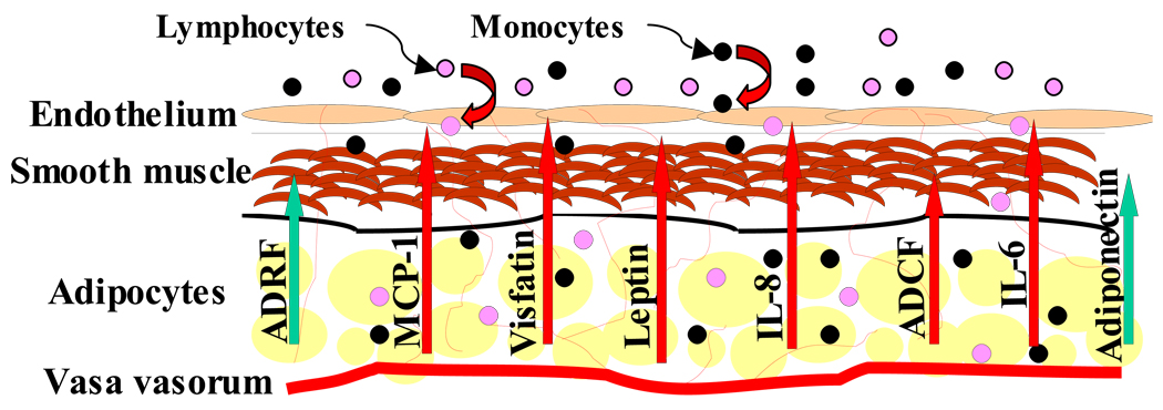
Crosstalk between cells in the blood vessel wall is vital to normal vascular function and is perturbed in diseases such as atherosclerosis and hypertension. Perivascular adipocytes reside at the adventitial border of blood vessels but until recently were virtually ignored in studies of vascular function. However, perivascular adipocytes have been demonstrated to be powerful endocrine cells capable of responding to metabolic cues and transducing signals to adjacent blood vessels. Accordingly, crosstalk between perivascular adipose tissue (PVAT) and blood vessels is now being intensely examined. Emerging evidence suggests that PVAT regulates vascular function through numerous mechanisms, but evidence to date suggests modulation of three key aspects that are the focus of this review: inflammation, vasoreactivity, and smooth muscle cell proliferation.
Copyright 2009 Elsevier Ltd. All rights reserved.
Figures
References
-
-
Chatterjee TK, Stoll LL, Denning GM, Harrelson A, Blomkalns AL, Idelman G, Rothenberg FG, Neltner B, Romig-Martin SA, Dickson EW, Rudich S, Weintraub NL. Proinflammatory phenotype of perivascular adipocytes: influence of high-fat feeding. Circ Res. 2009;104:541–549. This paper highlighted the unique characteristics of human perivascular adipocytes and demonstrated the influence of high fat feeding on PVAT in mice.
-
-
- Iacobellis G, Sharma AM. Epicardial adipose tissue as new cardio-metabolic risk marker and potential therapeutic target in the metabolic syndrome. Curr Pharm Des. 2007;13:2180–2184. - PubMed
-
- Kwon HM, Sangiorgi G, Ritman EL, Lerman A, McKenna C, Virmani R, Edwards WD, Holmes DR, Schwartz RS. Adventitial vasa vasorum in balloon-injured coronary arteries: visualization and quantitation by a microscopic three-dimensional computed tomography technique. J Am Coll Cardiol. 1998;32:2072–2079. - PubMed
Publication types
MeSH terms
Grants and funding
LinkOut - more resources
Full Text Sources
Other Literature Sources
Medical
