. 2010 Apr 1;686(1-2):39-46.
doi: 10.1016/j.mrfmmm.2009.12.012. Epub 2010 Jan 7.

ATR preferentially interacts with common fragile site FRA3B and the binding requires its kinase activity in response to aphidicolin treatment

Affiliations

ATR preferentially interacts with common fragile site FRA3B and the binding requires its kinase activity in response to aphidicolin treatment

Cheng Wan et al. Mutat Res. .

Abstract

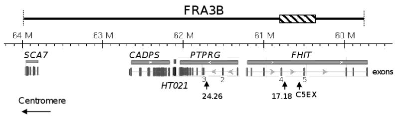
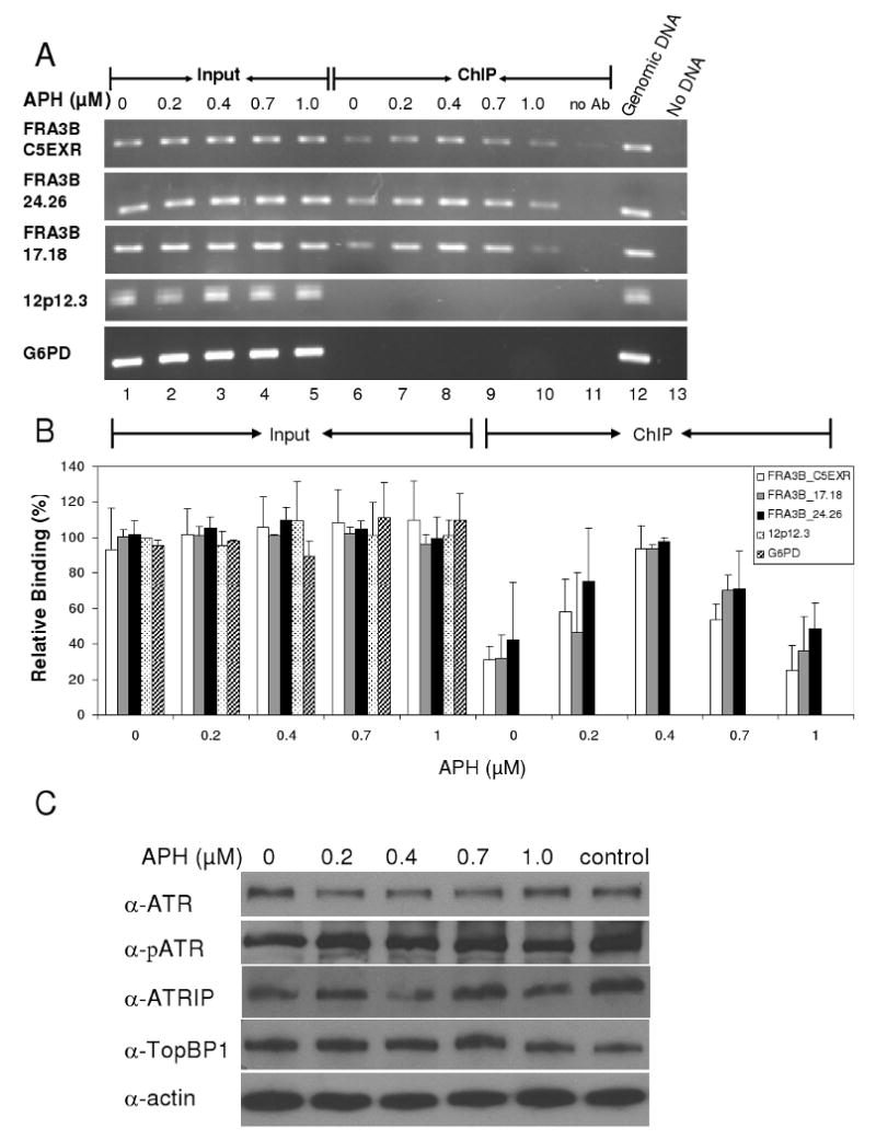
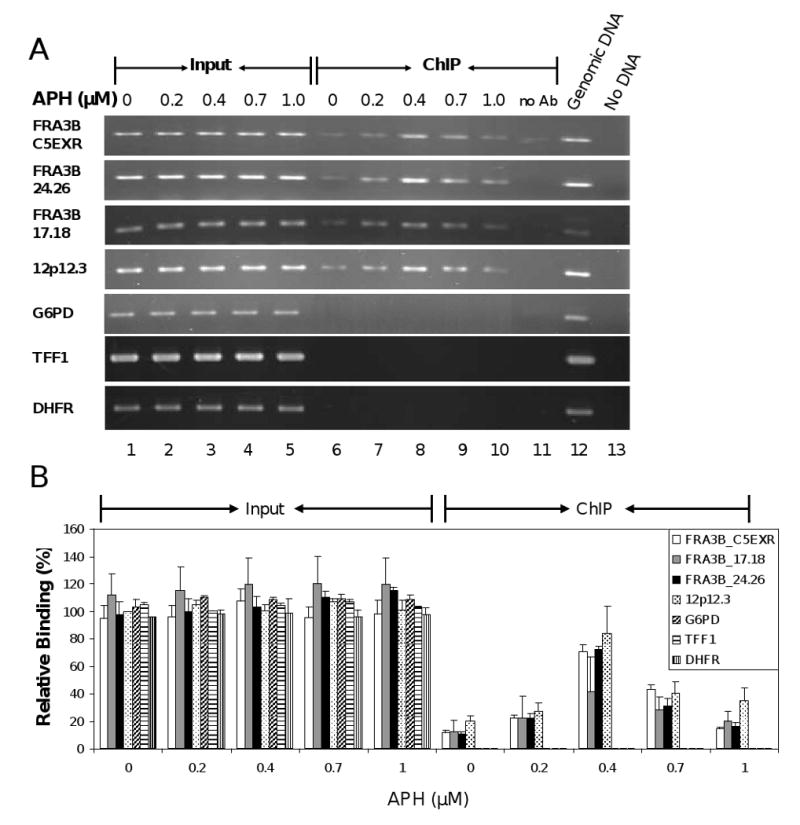
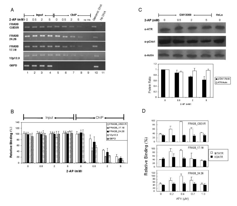
The instability of common fragile sites (CFSs) contributes to the development of a variety of cancers. The ATR-dependent DNA damage checkpoint pathway has been implicated in maintaining CFS stability, but the mechanism is incompletely understood. The goal of our study was to elucidate the action of the ATR protein in the CFS-specific ATR-dependent checkpoint response. Using a chromatin immunoprecipitation assay, we demonstrated that ATR protein preferentially binds (directly or through complexes) to fragile site FRA3B as compared to non-fragile site regions, under conditions of mild replication stress. Interestingly, the amount of ATR protein that bound to three regions of FRA3B peaked at 0.4microM aphidicolin (APH) treatment and decreased again at higher concentrations of APH. The total amounts of cellular ATR and several ATR-interacting proteins remained unchanged, suggesting that ATR binding to the fragile site is guided initially by the level of replication stress signals generated at FRA3B due to APH treatment and then sequestered from FRA3B regions by successive signals from other non-fragile site regions, which are produced at the higher concentrations of APH. This decrease in ATR binding to fragile site FRA3B at the higher concentrations of APH may account for the increasing number of chromosome gaps and breaks observed under the same conditions. Furthermore, inhibition of ATR kinase activity by treatment with 2-aminopurine (2-AP) or by over-expression of a kinase-dead ATR mutant showed that the kinase activity is required for the binding of ATR to fragile DNAs in response to APH treatment. Our results provide novel insight into the mechanism for the regulation of fragile site stability by ATR.

Keywords: ATR; FRA3B; Fragile site.


Conflict of interest statement

Conflict of interest statement: The authors declare that there are no conflicts of interest.

Figures

Fig. 1
Fig. 1
Genomic organization of the FRA3B region. Although the boundaries of FRA3B are not well defined, APH-induced fragility of FRA3B has been reported to span ∼4 Mb, including five genes - SCA7, CADPS, HT021, PTPRG, and FHIT [24]. The genomic coordinates (Human Genome GRch37 assembly) including distance scale are listed above each gene. Exons of each gene are listed as vertical bars below, and exons 4 and 5 of FHIT and exons 2 and 3 of PTPRG are numbered. The ∼500 kb unstable region of FRA3B which has been investigated thoroughly [12,13,25], extends from intron 3 (near exon 4) to intron 5 (∼150 kb distal to exon 5) of the FHIT gene, and is indicated by a hatched bar. The positions of the three examined regions: FRA3B_C5EXR (chr3:60516490-60516900), FRA3B_24.26 (chr3: 61742231-61742422), and FRA3B_17.18 (chr3:60719769-60719981) are indicated by vertical arrowheads.
Fig. 2
Fig. 2
ATR protein preferentially binds to FRA3B fragile site in HeLa cells upon APH treatment. (A) A representative PCR analysis of DNA immunoprecipitated from HeLa cells with anti-ATR antibody. HeLa cells were treated with various APH concentrations (0, 0.2, 0.4, 0.7, and 1.0 μM). The 0.1% of input DNAs (lanes 1-5) and chromatin immunoprecipitated (ChIP) DNAs (lanes 6-10) were amplified by PCR at the following DNA regions: FRA3B_C5EXR, FRA3B_24.26, FRA3B_17.18, 12p12.3, and G6PD. For each ChIP assay, a no-antibody control (lane 11) was included. For each PCR reaction, there was a PCR positive control in which 20 ng genomic DNA served as PCR template (lane 12) and a PCR negative control in which no DNA template was added (lane 13). (B) Quantitation of the level of ATR protein binding to various DNA regions in HeLa cells. The amount of DNA in either input or ChIP samples, relative to that of the input of the untreated 12p12.3 sample (which was assigned a value of 1), is an average from three separate but identical experiments. Average with standard deviation is shown. (C) The amount of total cellular ATR, phosphorylated ATR, ATRIP, TopBP1 proteins during APH induction. Whole cell lysates from HeLa cells treated with various amounts of APH were separated by SDS-PAGE, and analyzed with the respective antibodies and an anti-actin antibody.
Fig. 3
Fig. 3
ATR protein binds FRA3B fragile site in GM13069 cells upon APH treatment. (A) A representative PCR analysis of DNA immunoprecipitated from GM13069 cells with anti-ATR antibody. GM13069 cells were treated with various APH concentrations (0, 0.2, 0.4, 0.7, and 1.0 μM). The 0.1% of input DNAs (lanes 1-5) and chromatin immunoprecipitated DNAs (lanes 6-10) were amplified by PCR at the following DNA regions: FRA3B_C5EXR, FRA3B_24.26, FRA3B_17.18, 12p12.3, G6PD, TFF1 and DHFR. For each ChIP assay, a no-antibody control (lane 11) was included. For each PCR reaction, there was a PCR positive control in which 20 ng genomic DNA served as PCR template (lane 12) and a PCR negative control in which no DNA template was added (lane 13). (B) Quantitation of the level of ATR protein binding to various DNA regions in GM13069 cells. The amount of DNA in either input or ChIP samples, relative to that of the input of the untreated 12p12.3 sample (which was assigned a value of 1), is an average from four separate but identical experiments. Average with standard deviation is shown.
Fig. 4
Fig. 4
Kinase activity of ATR protein is required for binding to fragile DNA in response to APH treatment. (A) A representative PCR analysis of DNAs immunoprecipitated from GM13069 cells with anti-ATR antibody. GM13069 cells were treated with various 2-AP concentrations (0, 1, 2, and 5 mM), an inhibitor of ATR kinase activity, along with 0.4 μM APH. The 0.1 % of input DNA (lanes 1-4) and chromatin immunoprecipitated DNA (lanes 5-8) were amplified by PCR at the following DNA regions: FRA3B_C5EXR, FRA3B_24.26, FRA3B_17.18, 12p12.3, and G6PD. For each ChIP assay, a no-antibody control (lane 9) was included. For each PCR reaction, there was a PCR positive control in which 20 ng genomic DNA served as PCR template (lane 10) and a PCR negative control in which no DNA template was added (lane 11). (B) Quantitation of the level of ATR protein binding to various DNA regions in GM13069 cells. The amount of DNA in either input or ChIP samples, relative to that of the input of the untreated 12p12.3 sample (which was assigned a value of 1), is an average from four separate but identical experiments. Average with standard deviation is shown. (C) The amount of total cellular ATR protein during 2-AP treatment. Whole cell lysates from GM13069 cells, that were treated with various 2-AP concentrations along with 0.4 μM APH, were separated by SDS-PAGE, and analyzed with anti-ATR, anti-phospho-Chk1 (Ser345), or anti-actin antibodies. Quantitative analysis of protein ratios for ATR/actin (white bar) and p-Chk1/actin (black bar) was plotted. Average with standard deviation is shown. (D) ATR protein binding to three FRA3B regions in HeLa cells transfected with FLAG-tagged wild type (open bars) or kinase-dead (black bars) ATR constructs. Thirty hours after transfection, the cells were treated with APH (0, 0.2, 0.4, 0.7, and 1.0 μM) for an additional 24 h. ChIP assays were carried out with anti-FLAG antibody, and chromatin immunoprecipitated DNAs were amplified by PCR at the FRA3B_C5EXR, FRA3B_24.26, FRA3B_17.18, and 12p12.3 regions. The amount of DNA in ChIP samples, relative to the DNA input of the 12p12.3 sample from untreated, wild-type ATR transfected cells (which was assigned a value of 1), is an average of three separate but identical experiments. Average with standard deviation is shown. Both types of ATR showed no binding to the non-fragile 12p12.3 region (data not shown).

Similar articles

Cited by

References

    1. Casper AM, Nghiem P, Arlt MF, Glover TW. ATR regulates fragile site stability. Cell. 2002;111:779–789. - PubMed
    1. Casper AM, Durkin SG, Arlt MF, Glover TW. Chromosomal instability at common fragile sites in Seckel syndrome. Am J Hum Genet. 2004;75:654–660. - PMC - PubMed
    1. Arlt MF, Xu B, Durkin SG, Casper AM, Kastan MB, Glover TW. BRCA1 is required for common-fragile-site stability via its G2/M checkpoint function. Mol Cell Biol. 2004;24:6701–6709. - PMC - PubMed
    1. Durkin SG, Arlt MF, Howlett NG, Glover TW. Depletion of CHK1, but not CHK2, induces chromosomal instability and breaks at common fragile sites. Oncogene. 2006;25:4381–4388. - PubMed
    1. Howlett NG, Taniguchi T, Durkin SG, D'Andrea AD, Glover TW. The Fanconi anemia pathway is required for the DNA replication stress response and for the regulation of common fragile site stability. Hum Mol Genet. 2005;14:693–701. - PubMed

Publication types

MeSH terms

LinkOut - more resources