On the biomechanical function of scaffolds for engineering load-bearing soft tissues
- PMID: 20060509
- PMCID: PMC2878661
- DOI: 10.1016/j.actbio.2010.01.001
On the biomechanical function of scaffolds for engineering load-bearing soft tissues
Abstract
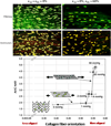
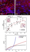
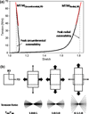
Replacement or regeneration of load-bearing soft tissues has long been the impetus for the development of bioactive materials. While maturing, current efforts continue to be confounded by our lack of understanding of the intricate multi-scale hierarchical arrangements and interactions typically found in native tissues. The current state of the art in biomaterial processing enables a degree of controllable microstructure that can be used for the development of model systems to deduce fundamental biological implications of matrix morphologies on cell function. Furthermore, the development of computational frameworks which allow for the simulation of experimentally derived observations represents a positive departure from what has mostly been an empirically driven field, enabling a deeper understanding of the highly complex biological mechanisms we wish to ultimately emulate. Ongoing research is actively pursuing new materials and processing methods to control material structure down to the micro-scale to sustain or improve cell viability, guide tissue growth, and provide mechanical integrity, all while exhibiting the capacity to degrade in a controlled manner. The purpose of this review is not to focus solely on material processing but to assess the ability of these techniques to produce mechanically sound tissue surrogates, highlight the unique structural characteristics produced in these materials, and discuss how this translates to distinct macroscopic biomechanical behaviors.
Copyright 2010 Acta Materialia Inc. Published by Elsevier Ltd. All rights reserved.
Figures


Similar articles
-
Design and analysis of tissue engineering scaffolds that mimic soft tissue mechanical anisotropy.Biomaterials. 2006 Jul;27(19):3631-8. doi: 10.1016/j.biomaterials.2006.02.024. Epub 2006 Mar 20. Biomaterials. 2006. PMID: 16545867
-
Hierarchical mechanics of connective tissues: integrating insights from nano to macroscopic studies.J Biomed Nanotechnol. 2014 Oct;10(10):2464-507. J Biomed Nanotechnol. 2014. PMID: 25992406 Review.
-
Electrospun nanofibrous scaffolds for engineering soft connective tissues.Methods Mol Biol. 2011;726:243-58. doi: 10.1007/978-1-61779-052-2_16. Methods Mol Biol. 2011. PMID: 21424454
-
Fabrication of dense anisotropic collagen scaffolds using biaxial compression.Acta Biomater. 2018 Jan;65:76-87. doi: 10.1016/j.actbio.2017.11.017. Epub 2017 Nov 8. Acta Biomater. 2018. PMID: 29128533 Free PMC article.
-
Factors influencing the long-term behavior of extracellular matrix-derived scaffolds for musculoskeletal soft tissue repair.J Long Term Eff Med Implants. 2012;22(3):181-93. doi: 10.1615/jlongtermeffmedimplants.2013006120. J Long Term Eff Med Implants. 2012. PMID: 23582110 Free PMC article. Review.
Cited by
-
The rationale and emergence of electroconductive biomaterial scaffolds in cardiac tissue engineering.APL Bioeng. 2019 Oct 15;3(4):041501. doi: 10.1063/1.5116579. eCollection 2019 Dec. APL Bioeng. 2019. PMID: 31650097 Free PMC article. Review.
-
From single fiber to macro-level mechanics: A structural finite-element model for elastomeric fibrous biomaterials.J Mech Behav Biomed Mater. 2014 Nov;39:146-61. doi: 10.1016/j.jmbbm.2014.07.016. Epub 2014 Aug 1. J Mech Behav Biomed Mater. 2014. PMID: 25128869 Free PMC article.
-
Establishing the Basis for Mechanobiology-Based Physical Therapy Protocols to Potentiate Cellular Healing and Tissue Regeneration.Front Physiol. 2017 Jun 6;8:303. doi: 10.3389/fphys.2017.00303. eCollection 2017. Front Physiol. 2017. PMID: 28634452 Free PMC article. Review.
-
How to make a heart valve: from embryonic development to bioengineering of living valve substitutes.Cold Spring Harb Perspect Med. 2014 Nov 3;4(11):a013912. doi: 10.1101/cshperspect.a013912. Cold Spring Harb Perspect Med. 2014. PMID: 25368013 Free PMC article. Review.
-
Improved cellular infiltration in electrospun fiber via engineered porosity.Tissue Eng. 2007 Sep;13(9):2249-57. doi: 10.1089/ten.2006.0306. Tissue Eng. 2007. PMID: 17536926 Free PMC article.
References
-
- Gilbert TW, et al. Degradation and remodeling of small intestinal submucosa in canine Achilles tendon repair. J Bone Joint Surg Am. 2007;89(3):621–630. - PubMed
-
- Shelton JC, Bader DL, Lee DA. Mechanical conditioning influences the metabolic response of cell-seeded constructs. Cells Tissues Organs. 2003;175(3):140–150. - PubMed
-
- van der Meulen MC, Huiskes R. Why mechanobiology? A survey article. J Biomech. 2002;35(4):401–414. - PubMed
-
- Sarkadi B, Parker JC. Activation of ion transport pathways by changes in cell volume. Biochim Biophys Acta. 1991;1071(4):407–427. - PubMed
Publication types
MeSH terms
Grants and funding
LinkOut - more resources
Full Text Sources
Other Literature Sources

