Review
. 2010 Mar;33(3):130-9.
doi: 10.1016/j.tins.2009.12.004. Epub 2010 Jan 8.

Early transformations in odor representation

Affiliations
Review

Early transformations in odor representation

Thomas A Cleland. Trends Neurosci. 2010 Mar.

Abstract

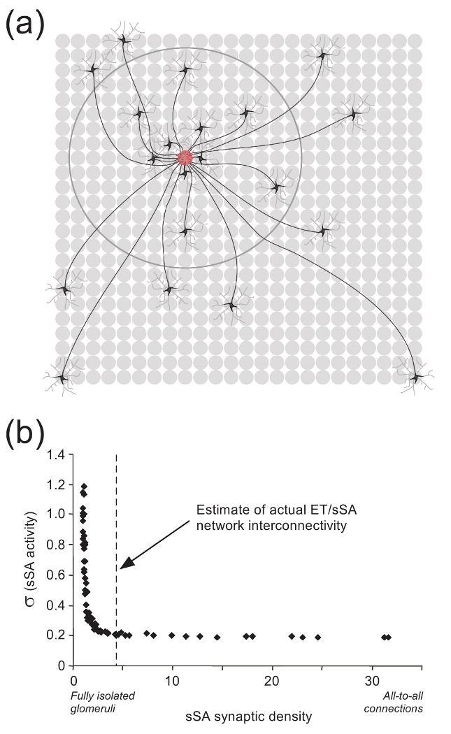
Sensory representations are repeatedly transformed by neural computations that determine which of their attributes can be effectively processed at each stage. Whereas some early computations are common across multiple sensory systems, they can utilize dissimilar underlying mechanisms depending on the properties of each modality. Recent work in the olfactory bulb has substantially clarified the neural algorithms underlying early odor processing. The high-dimensionality of odor space strictly limits the utility of topographical representations, forcing similarity-dependent computations such as decorrelation to employ unusual neural algorithms. The distinct architectures and properties of the two prominent computational layers in the olfactory bulb suggest that the bulb is directly comparable not only to the retina but also to primary visual cortex.


Figures

Figure 1
Figure 1
(a) Circuit diagram of the mammalian olfactory bulb. The axons of olfactory sensory neurons expressing the same odorant receptor type converge together as they cross the cribriform plate and arborize together to form glomeruli (shaded ovals) across the surface of the olfactory bulb. Several classes of olfactory bulb neuron innervate each glomerulus, including both principal neurons and intrinsic interneurons. Glomerular interneuron classes are heterogeneous, including olfactory nerve-driven periglomerular cells (PGo), external tufted cell-driven periglomerular cells (PGe), and multiple subtypes of external tufted cells (ET). Superficial short-axon cells (sSA) are not associated with specific glomeruli but project broadly and laterally within the deep glomerular layer, interacting with glomerular interneurons. Principal neurons include mitral cells (Mi), which interact via reciprocal connections in the external plexiform layer (EPL) with the dendrites of inhibitory granule cells (Gr), thereby receiving recurrent and lateral inhibition. Middle/deep tufted cells (not depicted) constitute another, less understood class of olfactory bulb principal neurons noted for their relative lack of an inhibitory surround. Both of these principal neuron types project divergently to several regions of the brain, though the projection profiles of the two classes differ [71, 90]. A sparse, heterogeneous population of inhibitory interneurons known collectively as deep short-axon cells [91] also is not depicted. OE, olfactory epithelium (in the nasal cavity); GL, glomerular layer; EPL; external plexiform layer; MCL, mitral cell layer; IPL, internal plexiform layer; GCL, granule cell layer. Filled triangles denote excitatory (glutamatergic) synapses; open circles denote inhibitory (GABAergic) synapses. (b) Schematic depiction of decorrelation between two overlapping representations (α and β), depicted in one dimension (left panel). Canonical “Mexican-hat” decorrelation (upper right panel) generates an explicit inhibitory surround in which the edges of the representation are inhibited below baseline, yielding a sharp reduction in overlap among similar representations. This computation is performed by lateral inhibition in the retina and inferior colliculus, and by the nontopographical model of olfactory receptive field decorrelation. A lesser degree of decorrelation can also be obtained by broad, nonspecific inhibition, including lateral inhibition with an unstructured surround [45, 46] (lower right panel), although this imposes a general reduction in activity across the entire representation. This operation is the general result of lateral inhibitory mechanisms as studied to date in the olfactory bulb. While both computations can effect a measurable decorrelation, the two transformations differ substantially. (c) Replication of experimental data from [23] by the nontopographical model [26] demonstrating Mexican-hat decorrelation in mitral cells among the responses to similar odorants (3-carbon through 11-carbon aliphatic aldehydes). Periodic bursts of spikes reflect background activity evoked by the respiration cycle; a 2 second odorant stimulus was presented during the third inhalation (black bar; green shading). The odorant hexanal ((6)CHO) is near the center of this mitral cell's receptive field and evokes the strongest activation; pentanal and heptanal also excite the cell, whereas butanal ((4)CHO) and octanal ((5)CHO) are within its inhibitory surround, and hence evoke a net inhibition. The mitral cell is unresponsive to the other four odorants. The right panel illustrates how the Mexican-hat function maps onto the trajectory through odor similarity space defined by the homologous odor series. Plus sign connotes excitation; minus sign connotes inhibition. Figure adapted from ref. [26].
Figure 2
Figure 2
Illustration of the nontopographical model for olfactory receptive field decorrelation. (a) Level of activation of selected MOB neurons as a function of ligand-receptor potency in its presynaptic OSN population. All response profiles depicted are from neurons innervating the same glomerulus. Negative values of neuronal activation connote inhibition. Odors with very weak potencies for the OR in question evoke no OSN activity and hence no mitral cell activity. Increasing the ligand-receptor potency to the point where it evokes OSN activity begins to excite PGo neurons, which owing to their high input resistance and small gemmule volume respond strongly to even weak inputs and deliver local intraglomerular inhibition onto mitral cells. Moderate ligand-receptor potencies begin to also directly activate mitral cells (Miin), but this excitation is overpowered by the inhibition received from the more strongly activated PGo neurons, which shunt away depolarizing current such that the overall net response of mitral cells (Miout) is inhibitory. Strong ligand-receptor potencies excite mitral cells more strongly, overwhelming the capacity of PGo inhibition to impair spike generation and hence evoking action potentials in mitral cells. The result is that mitral cells exhibit an excitatory response to high-potency odorant ligands, and an inhibitory response to odorant ligands of moderate potency – i.e., to the “surrounding” region in a space defined by odor quality, as illustrated in Figure 1c. Figure adapted from ref. [26]. (b) Illustration of the triune synapse at which an OSN excites a mitral cell and PGo cell gemmule in parallel, and the PGo cell immediately inhibits the mitral cell. This synaptic triad is the basis for nontopographical intraglomerular inhibition proposed to mediate decorrelation among similar OR receptive fields.
Figure 3
Figure 3
Normalization of mitral cell gross activity levels via the external tufted/superficial short-axon cell (ET/sSA) network. (a) Schematic depiction of lateral connectivity in the deep glomerular layer via sSA cells [48]. Dye injections into single glomeruli (red glomerulus) indicate that ∼50 sSA neurons project axons to a given glomerulus (twenty sSA neurons are depicted here); furthermore, whereas sSA cell axons branch extensively, for clarity only one axonal branch per sSA neuron is depicted here. The dendritic arbors of sSA neurons extend across a small number of glomeruli (depicted as light grey arbors around each sSA neuron). Of the sSA neurons projecting axons to a given glomerulus, 50% are located over 5-7 glomerular diameters away from the injected glomerulus (denoted by large circle), whereas 10% are located over 15-18 glomerular diameters distant (two neurons depicted in lower corners). The longest sSA axons extend 20-30 glomerular diameters, an appreciable fraction of the circumference of the MOB (∼80 glomerular diameters in rats or mice). Grey circles denote glomeruli. Data drawn from ref. [48]. (b) The interglomerular connectivity of the ET/sSA network is functionally equivalent to a fully-connected all-to-all network [25]. The abscissa denotes an overall measure of sSA network connectivity, between the hypothetical extremes of no ET/sSA connectivity at all (fully isolated glomeruli) and full connectivity in which every glomerulus is directly linked to every other glomerulus (all-to-all connections). The greater the connectivity, the lower the variance in the activation levels among sSA neurons. That is, zero connectivity means that sSA neurons directly inherit (via ET cells) the heterogeneous odor-evoked activation levels of the OSNs associated with the nearest glomerulus, such that different sSA neurons differ widely in their activation levels across the MOB. In contrast, full connectivity implies that every sSA neuron receives essentially the same amount of afferent input (by receiving excitation drawn from every glomerulus in the MOB), which produces minimal variance in the activity levels among different sSA neurons. An estimate of actual ET/sSA connectivity in the mouse MOB (dashed vertical line) suggests that this center-surround connectivity pattern exerts the same quantitative, normalizing effect as would a fully-connected all-to-all network, but at a fraction of the metabolic cost. Figure adapted from ref. [25].

References

    1. Marr D. Vision: A computational investigation into the human representation and processing of visual information. W. H. Freeman; 1983.
    1. Marr D, Poggio T. MIT AI Memo #357. 1976. From understanding computation to understanding neural circuitry.
    1. Kleene SJ. The electrochemical basis of odor transduction in vertebrate olfactory cilia. Chem Senses. 2008;33:839–859. - PubMed
    1. Serizawa S, et al. Negative feedback regulation ensures the one receptor-one olfactory neuron rule in mouse. Science. 2003;302:2088–2094. - PubMed
    1. Nguyen MQ, et al. Prominent roles for odorant receptor coding sequences in allelic exclusion. Cell. 2007;131:1009–1017. - PMC - PubMed

Publication types