

Collapsibility of lung volume by paired inspiratory and expiratory CT scans: correlations with lung function and mean lung density
- PMID: 20060751
- PMCID: PMC2834821
- DOI: 10.1016/j.acra.2009.11.004
Collapsibility of lung volume by paired inspiratory and expiratory CT scans: correlations with lung function and mean lung density
Abstract
Rationale and objectives: To evaluate the relationship between measurements of lung volume (LV) on inspiratory/expiratory computed tomography (CT) scans, pulmonary function tests (PFT), and CT measurements of emphysema in individuals with chronic obstructive pulmonary disease.
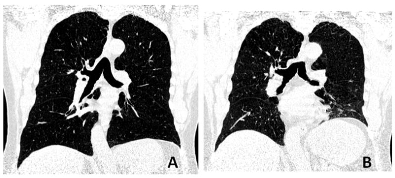
Materials and methods: Forty-six smokers (20 females and 26 males; age range 46-81 years), enrolled in the Lung Tissue Research Consortium, underwent PFT and chest CT at full inspiration and expiration. Inspiratory and expiratory LV values were automatically measured by open-source software, and the expiratory/inspiratory (E/I) ratio of LV was calculated. Mean lung density (MLD) and low attenuation area percent (<-950 HU) were also measured. Correlations of LV measurements with lung function and other CT indices were evaluated by the Spearman rank correlation test.
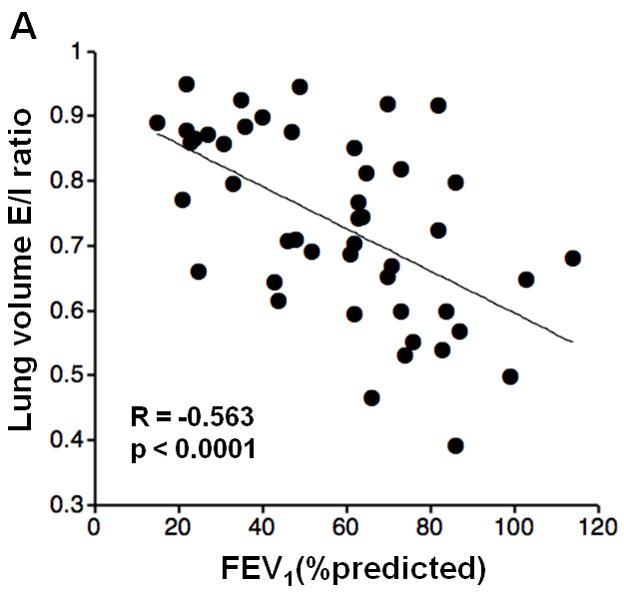
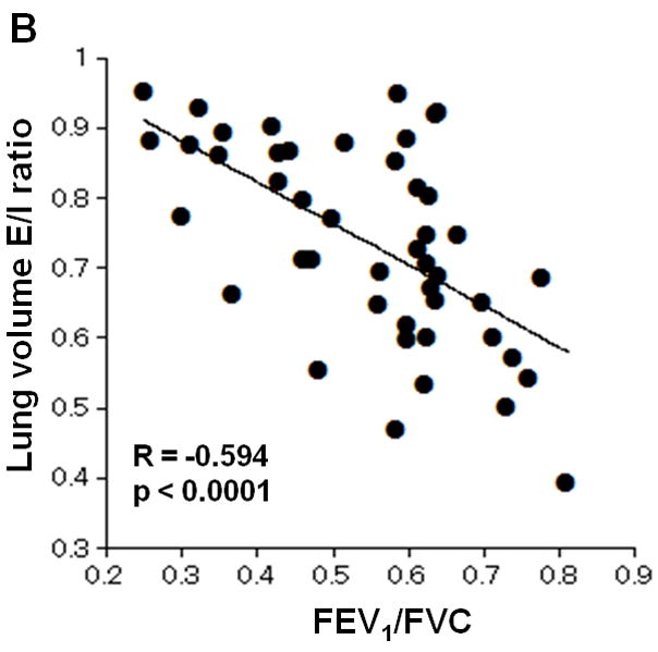
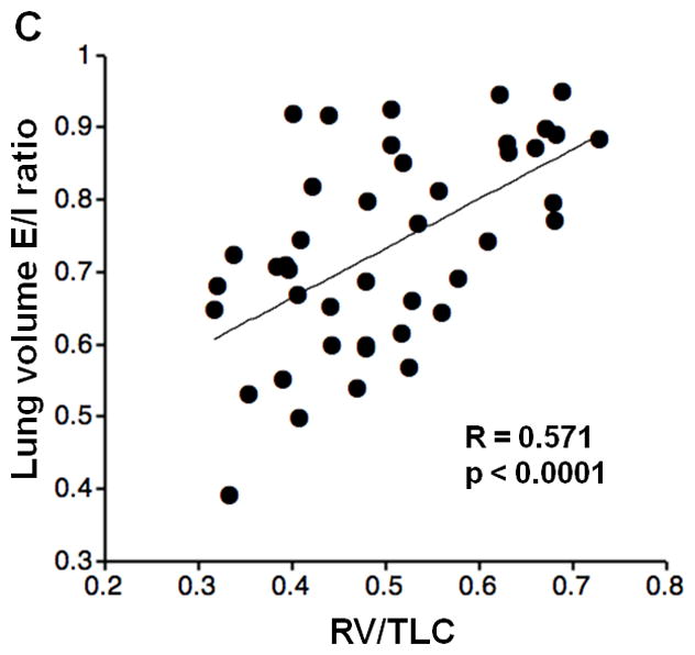
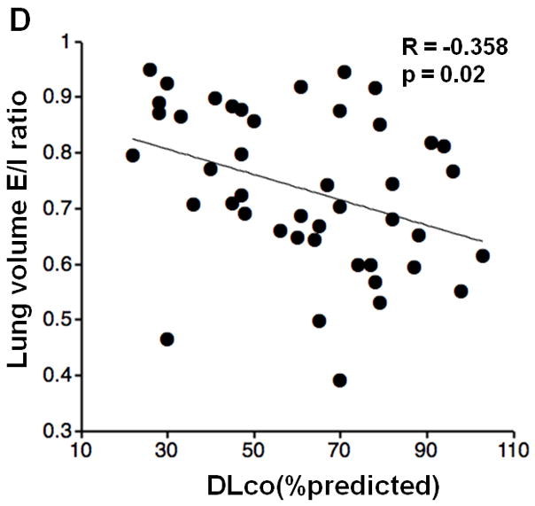
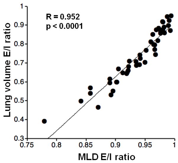
Results: LV E/I ratio significantly correlated with the following: the percentage of predicted value of forced expiratory volume in the first second (FEV(1)), the ratio of FEV(1) to forced vital capacity (FVC), and the ratio of residual volume (RV) to total lung capacity (TLC) (FEV(1)%P, R = -0.56, P < .0001; FEV(1)/FVC, r = -0.59, P < .0001; RV/TLC, r = 0.57, P < .0001, respectively). A higher correlation coefficient was observed between expiratory LV and expiratory MLD (r = -0.73, P < .0001) than between inspiratory LV and inspiratory MLD (r = -0.46, P < .01). LV E/I ratio showed a very strong correlation to MLD E/I ratio (r = 0.95, P < .0001).
Conclusions: LV E/I ratio can be considered to be equivalent to MLD E/I ratio and to reflect airflow limitation and air-trapping. Higher collapsibility of lung volume, observed by inspiratory/expiratory CT, indicates less severe conditions in chronic obstructive pulmonary disease.
Keywords: airflow obstruction; chronic obstructive pulmonary disease; computed tomography; lung volume; pulmonary emphysema.
Copyright 2010 AUR. Published by Elsevier Inc. All rights reserved.
Conflict of interest statement
Figures
Similar articles
-
Kurtosis and skewness of density histograms on inspiratory and expiratory CT scans in smokers.COPD. 2011 Feb;8(1):13-20. doi: 10.3109/15412555.2010.541537. COPD. 2011. PMID: 21299474
-
[Quantitative analysis of emphysema and air trapping at inspiratory and expiratory phase multi-slice spiral CT scan in smokers: correlation with pulmonary function test].Zhonghua Yi Xue Za Zhi. 2018 May 22;98(19):1467-1473. doi: 10.3760/cma.j.issn.0376-2491.2018.19.003. Zhonghua Yi Xue Za Zhi. 2018. PMID: 29804412 Chinese.
-
Quantitative CT in chronic obstructive pulmonary disease: inspiratory and expiratory assessment.AJR Am J Roentgenol. 2009 Jan;192(1):267-72. doi: 10.2214/AJR.07.3953. AJR Am J Roentgenol. 2009. PMID: 19098209
-
Paired inspiratory/expiratory volumetric thin-slice CT scan for emphysema analysis: comparison of different quantitative evaluations and pulmonary function test.Chest. 2005 Nov;128(5):3212-20. doi: 10.1378/chest.128.5.3212. Chest. 2005. PMID: 16304264
-
Quantitative computed tomography measurements to evaluate airway disease in chronic obstructive pulmonary disease: Relationship to physiological measurements, clinical index and visual assessment of airway disease.Eur J Radiol. 2016 Nov;85(11):2144-2151. doi: 10.1016/j.ejrad.2016.09.010. Epub 2016 Sep 13. Eur J Radiol. 2016. PMID: 27776670 Free PMC article. Review.
Cited by
-
Acute administration of ivacaftor to people with cystic fibrosis and a G551D-CFTR mutation reveals smooth muscle abnormalities.JCI Insight. 2016 Apr 7;1(4):e86183. doi: 10.1172/jci.insight.86183. JCI Insight. 2016. PMID: 27158673 Free PMC article.
-
Continuous quantitative measurement of the main bronchial dimensions and lung density in the lateral position by four-dimensional dynamic-ventilation CT in smokers and COPD patients.Int J Chron Obstruct Pulmon Dis. 2018 Nov 27;13:3845-3856. doi: 10.2147/COPD.S178836. eCollection 2018. Int J Chron Obstruct Pulmon Dis. 2018. PMID: 30568436 Free PMC article.
-
Detailed analysis of the density change on chest CT of COPD using non-rigid registration of inspiration/expiration CT scans.Eur Radiol. 2015 Feb;25(2):541-9. doi: 10.1007/s00330-014-3418-0. Epub 2014 Sep 14. Eur Radiol. 2015. PMID: 25218764
-
Lobar analysis of collapsibility indices to assess functional lung volumes in COPD patients.Int J Chron Obstruct Pulmon Dis. 2014 Dec 9;9:1347-56. doi: 10.2147/COPD.S72616. eCollection 2014. Int J Chron Obstruct Pulmon Dis. 2014. PMID: 25525352 Free PMC article.
-
Automated continuous quantitative measurement of proximal airways on dynamic ventilation CT: initial experience using an ex vivo porcine lung phantom.Int J Chron Obstruct Pulmon Dis. 2015 Sep 25;10:2045-54. doi: 10.2147/COPD.S87588. eCollection 2015. Int J Chron Obstruct Pulmon Dis. 2015. PMID: 26445535 Free PMC article.
References
-
- Kauczor HU, Heussel CP, Fischer B, Klamm R, Mildenberger P, Thelen M. Assessment of lung volumes using helical CT at inspiration and expiration: comparison with pulmonary function tests. Am J Roentgenol. 1998;171:1091–1095. - PubMed
-
- Zaporozhan J, Ley S, Eberhardt R, Weinheimer O, Iliyushenko S, Herth F, et al. Paired inspiratory/expiratory volumetric thin-section CT scan for emphysema analysis: comparison of different quantitative evaluations and pulmonary function test. Chest. 2005;128:3212–3220. - PubMed
-
- Akira M, Toyokawa K, Inoue Y, Arai T. Quantitative CT in chronic obstructive pulmonary disease: inspiratory and expiratory assessment. Am J Roentgenol. 2009;192:267–272. - PubMed
-
- Lee YK, Oh YM, Lee JH, Kim EK, Lee JH, Kim N, et al. Quantitative assessment of emphysema, air trapping, and airway thickening on computed tomography. Lung. 2008;186:157–165. - PubMed
Publication types
MeSH terms
Grants and funding
LinkOut - more resources
Full Text Sources
Other Literature Sources
Medical
