. 2010 Apr 15;316(7):1254-62.
doi: 10.1016/j.yexcr.2010.01.007. Epub 2010 Jan 11.

Up- or downregulation of tescalcin in HL-60 cells is associated with their differentiation to either granulocytic or macrophage-like lineage

Affiliations

Up- or downregulation of tescalcin in HL-60 cells is associated with their differentiation to either granulocytic or macrophage-like lineage

Konstantin Levay et al. Exp Cell Res. .

Abstract

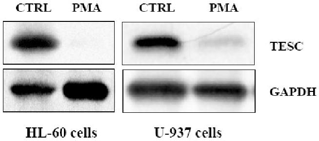
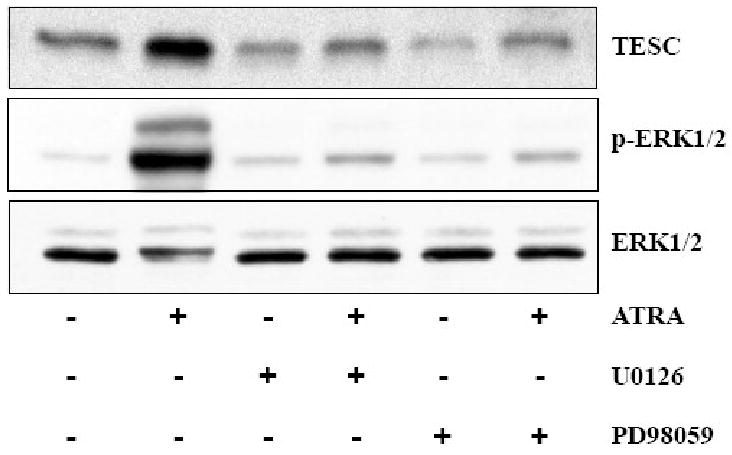
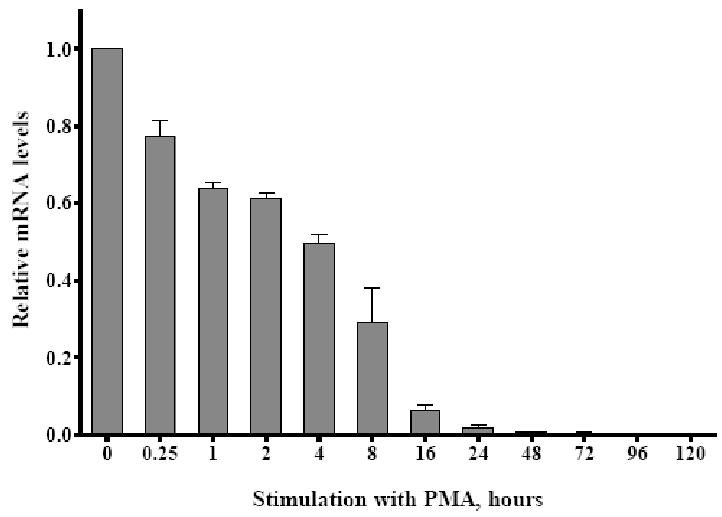
Tescalcin is a 25-kDa EF-hand Ca(2+)-binding protein that is differentially expressed in several mammalian tissues. Previous studies demonstrated that expression of this protein is essential for differentiation of hematopoietic precursor cell lines and primary stem cells into megakaryocytes. Here we show that tescalcin is expressed in primary human granulocytes and is upregulated in human promyelocytic leukemia HL-60 cells that have been induced to differentiate along the granulocytic lineage. However, during induced macrophage-like differentiation of HL-60 cells the expression of tescalcin is downregulated. The decrease in expression is associated with a rapid drop in tescalcin mRNA level, whereas upregulation occurs via a post-transcriptional mechanism. Tescalcin is necessary for HL-60 differentiation into granulocytes as its knockdown by shRNA impairs the ability of HL-60 cells to acquire the characteristic phenotypes such as phagocytic activity and generation of reactive oxygen species measured by respiratory burst assay. Both up- and downregulation of tescalcin require activation of the MEK/ERK cascade. It appears that commitment of HL-60 cells toward granulocytic versus macrophage-like lineage correlates with expression of tescalcin and kinetics of ERK activation. In retinoic acid-induced granulocytic differentiation, the activation of ERK and upregulation of tescalcin occurs slowly (16-48 h). In contrast, in PMA-induced macrophage-like differentiation the activation of ERK is rapid (15-30 min) and tescalcin is downregulated. These studies indicate that tescalcin is one of the key gene products that is involved in switching differentiation program in some cell types.


Figures

Figure 1
Figure 1. Expression of tescalcin during granulocytic differentiation of HL-60 cells and in primary granulocytes
(A) HL-60 cells were cultured as described in Materials and Methods and treated for 5 days either with 1mM ATRA, 1.25% DMSO, 80 mM DMF to induce granulocytic differentiation or with 0.1% DMSO vehicle as a control (CTRL). Cells were harvested in a lysis buffer on ice and approximately 15 μg of total proteins were analyzed by Western blot with antibody against tescalcin. Nitrocellulose membranes were then stripped and re-probed with antibody against GAPDH as a loading control. (B) Human polymorphonuclear granulocytes were isolated from peripheral blood as described in Material and Methods. Total protein extract (15 μg) was resolved on 10-20% gradient polyacrylamide gel (Novex) and analyzed by Western blot with antibody against tescalcin.
Figure 2
Figure 2. Downregulation of tescalcin during monocyte/macrophage differentiation of HL-60 and U-937 cells
To induce monocyte/macrophage-like differentiation, HL-60 or U-937 cells were treated for 5 days with 16 nM PMA or with 0.1% DMSO vehicle (CTRL). The level of tescalcin in total cell lysates was analyzed by Western blot with antibody against tescalcin. Antibody against GAPDH was used to show equal protein loading. Approximately 60 μg of total protein was loaded.
Figure 3
Figure 3. Time-course of tescalcin upregulation and activation of ERK during granulocytic differentiation of HL-60 cells
Exponentially growing HL-60 cells were stimulated with 1μM ATRA to induce granulocytic differentiation. Samples were collected at indicated times and analyzed by immunoblotting with antibodies against tescalcin, total p42/44 (ERK1/2) and phosphorylated p42/44 (p-ERK1/2). Antibody against β-actin was used to demonstrate equal protein loading.
Figure 4
Figure 4. ATRA-induced tescalcin upregulation requires activation of ERK
MEK inhibitors U0126 (final concentration in the medium 4 μM) or PD98059 (10 μM) were added to cultured HL-60 cells for 30 min prior to induction of granulocytic differentiation with ATRA. Cells were harvested 72 h. later, lysed on ice and analyzed by immunoblotting with antibodies against tescalcin, phosphorylated p42/44 (p-ERK1/2) and total p42/44 (ERK1/2).
Figure 5
Figure 5. Tescalcin gene expression in HL-60 cells is not affected by ATRA
Exponentially growing cells were treated with 1μM ATRA to initiate granulocytic differentiation. At the indicated time points cells were collected and total RNA was purified using RNeasy kit (Qiagen). Total RNA was transcribed with High Capacity cDNA Reverse Transcription Kit (Applied Biosystems). The relative expression levels of mRNAs encoding tescalcin, CD38, and c-Myc were analyzed by RQ-PCR and specific primers and probes (Taqman, Applied Biosystems). Reactions were run in triplicates on 7900HT Fast Real Time PCR system (Applied Biosystems) and normalized to the endogenous control 18S ribosomal RNA. Data are expressed as fold change relative to the gene expression levels at time 0 (mean±sd).
Figure 6
Figure 6. shRNA-mediated knockdown of tescalcin in HL-60 cells
(A) HL-60 cells were electroporated with plasmids encoding scrambled (CTRL) and Tescalcin-specific shRNA (shTESC), then selected for neomycin resistance. Expression of tescalcin in non-induced and ATRA-induced cells was detected by Western blot and compared to the wild-type HL-60. GAPDH is shown as loading control. (B) Total RNA was extracted from cells expressing either scrambled or tescalcin-specific shRNA and analyzed by RQ-PCR using specific primers and probes. Reactions were run in triplicates on 7900HT Fast Real Time PCR system (Applied Biosystems) and normalized to the endogenous control 18S ribosomal RNA. Data are expressed relative to wild-type HL-60 (mean±sd). Statistical analysis on results was performed using paired, two-tailed Student's t test and marked with a double asterisk if p < 0.01.
Figure 7
Figure 7. Tescalcin knockdown inhibits ATRA-induced granulocytic differentiation and maturation of HL-60 cells
Granulocytic differentiation was induced in HL-60 cells expressing scrambled or tescalcin-specific shRNA. (A) To measure the generation of superoxide, cells were collected at indicated times, washed with PBS, and incubated with 0.1% NBT as described in Material and Methods. The percentage of cells containing intracellular blue-black formazan deposits was then determined on Wright-Giemsa stained slide preparations. At least 400 cells were analyzed in each time point. (B) HL-60 cells were cultured with 1 μM ATRA or 0.1% DMSO (CTRL) for 72 hours. To study phagocytic activity, the cells were washed and incubated in fresh culture media with 0.025% of 1 μm fluorescent carboxylate-modified microspheres (Invitrogen). The fluorescence uptake was analyzed by flow cytometry as described under Material and Methods, with a minimum of 10,000 events acquired per sample. The number of cells ingested the fluorescent microspheres is expressed as % of total cells gated. Assays were performed in triplicates. Bar graph represents results of three independent experiments (mean±sd). Statistical analysis was performed using paired, two-tailed Student's t test.
Figure 8
Figure 8. Downregulation of tescalcin by PMA requires activation of ERK
HL-60 cells were cultured for 1 hour in the absence or presence of 4 μM U0126 MEK inhibitor prior to treatment with 16 nM PMA. Total protein extracts were prepared from cells harvested 72 hours later, analyzed by immunoblotting with tescalcin antibody, and compared to untreated HL-60. Antibody against GAPDH was used to show equal protein loading.
Figure 9
Figure 9. Kinetics of tescalcin downregulation and activation of ERK during PMA-induced macrophage-like differentiation
Cultured HL-60 cells were treated with 16 nM PMA to stimulate macrophage-like differentiation. Cells were harvested at the indicated times and total protein lysates were subjected to PAGE. Resolved proteins were analyzed by immunoblotting with antibodies against tescalcin, total p42/44 (ERK1/2) and phospho-p42/44 (p-ERK1/2).
Figure 10
Figure 10. PMA-mediated downregulation of tescalcin gene expression
To initiate macrophage-like differentiation, HL-60 cells were treated with 16 nM PMA. Total RNA was harvested from cells at the indicated time points, transcribed with High Capacity cDNA Reverse Transcription Kit and analyzed by RQ-PCR using tescalcin-specific primers and probes (Taqman, Applied Biosystems). Reactions were run in triplicates on 7900HT Fast Real Time PCR system (Applied Biosystems) and normalized to 18S rRNA. Data are expressed as fold change relative to the gene expression levels at time 0 (mean±sd).

Similar articles

Cited by

  • Regulation of Cop9 signalosome activity by the EF-hand Ca2+-binding protein tescalcin.
    Levay K, Slepak VZ. Levay K, et al. J Cell Sci. 2014 Jun 1;127(Pt 11):2448-59. doi: 10.1242/jcs.139592. Epub 2014 Mar 21. J Cell Sci. 2014. PMID: 24659803 Free PMC article.
  • Tescalcin/c-Src/IGF1Rβ-mediated STAT3 activation enhances cancer stemness and radioresistant properties through ALDH1.
    Lee JH, Choi SI, Kim RK, Cho EW, Kim IG. Lee JH, et al. Sci Rep. 2018 Jul 16;8(1):10711. doi: 10.1038/s41598-018-29142-x. Sci Rep. 2018. PMID: 30013043 Free PMC article.
  • Identification of common variants associated with human hippocampal and intracranial volumes.
    Stein JL, Medland SE, Vasquez AA, Hibar DP, Senstad RE, Winkler AM, Toro R, Appel K, Bartecek R, Bergmann Ø, Bernard M, Brown AA, Cannon DM, Chakravarty MM, Christoforou A, Domin M, Grimm O, Hollinshead M, Holmes AJ, Homuth G, Hottenga JJ, Langan C, Lopez LM, Hansell NK, Hwang KS, Kim S, Laje G, Lee PH, Liu X, Loth E, Lourdusamy A, Mattingsdal M, Mohnke S, Maniega SM, Nho K, Nugent AC, O'Brien C, Papmeyer M, Pütz B, Ramasamy A, Rasmussen J, Rijpkema M, Risacher SL, Roddey JC, Rose EJ, Ryten M, Shen L, Sprooten E, Strengman E, Teumer A, Trabzuni D, Turner J, van Eijk K, van Erp TG, van Tol MJ, Wittfeld K, Wolf C, Woudstra S, Aleman A, Alhusaini S, Almasy L, Binder EB, Brohawn DG, Cantor RM, Carless MA, Corvin A, Czisch M, Curran JE, Davies G, de Almeida MA, Delanty N, Depondt C, Duggirala R, Dyer TD, Erk S, Fagerness J, Fox PT, Freimer NB, Gill M, Göring HH, Hagler DJ, Hoehn D, Holsboer F, Hoogman M, Hosten N, Jahanshad N, Johnson MP, Kasperaviciute D, Kent JW Jr, Kochunov P, Lancaster JL, Lawrie SM, Liewald DC, Mandl R, Matarin M, Mattheisen M, Meisenzahl E, Melle I, Moses EK, Mühleisen TW, Nauck M, Nöthen MM, Olvera RL, Pandolfo M, Pike GB, Puls R, Reinvang I, Rentería ME, Rietsc… See abstract for full author list ➔ Stein JL, et al. Nat Genet. 2012 Apr 15;44(5):552-61. doi: 10.1038/ng.2250. Nat Genet. 2012. PMID: 22504417 Free PMC article.
  • TOX is required for development of the CD4 T cell lineage gene program.
    Aliahmad P, Kadavallore A, de la Torre B, Kappes D, Kaye J. Aliahmad P, et al. J Immunol. 2011 Dec 1;187(11):5931-40. doi: 10.4049/jimmunol.1101474. Epub 2011 Oct 21. J Immunol. 2011. PMID: 22021617 Free PMC article.
  • Genomic structural variants are linked with intellectual disability.
    Bulayeva K, Lesch KP, Bulayev O, Walsh C, Glatt S, Gurgenova F, Omarova J, Berdichevets I, Thompson PM. Bulayeva K, et al. J Neural Transm (Vienna). 2015 Sep;122(9):1289-301. doi: 10.1007/s00702-015-1366-8. Epub 2015 Jan 28. J Neural Transm (Vienna). 2015. PMID: 25626716 Free PMC article.

References

    1. Bennett JM, Catovsky D, Daniel MT, Flandrin G, Galton DA, Gralnick HR, Sultan C. Proposals for the classification of the acute leukaemias. French-American-British (FAB) co-operative group. Br J Haematol. 1976;33:451–458. - PubMed
    1. Dalton WT, Jr, Ahearn MJ, McCredie KB, Freireich EJ, Stass SA, Trujillo JM. HL-60 cell line was derived from a patient with FAB-M2 and not FAB-M3. Blood. 1988;71:242–247. - PubMed
    1. Breitman TR, Selonick SE, Collins SJ. Induction of differentiation of the human promyelocytic leukemia cell line (HL-60) by retinoic acid. Proc Natl Acad Sci U S A. 1980;77:2936–2940. - PMC - PubMed
    1. Collins SJ. The HL-60 promyelocytic leukemia cell line: proliferation, differentiation, and cellular oncogene expression. Blood. 1987;70:1233–1244. - PubMed
    1. Collins SJ, Ruscetti FW, Gallagher RE, Gallo RC. Terminal differentiation of human promyelocytic leukemia cells induced by dimethyl sulfoxide and other polar compounds. Proc Natl Acad Sci U S A. 1978;75:2458–2462. - PMC - PubMed

Publication types

MeSH terms