

The structure of the Salmonella typhimurium type III secretion system needle shows divergence from the flagellar system
- PMID: 20060835
- PMCID: PMC2823972
- DOI: 10.1016/j.jmb.2010.01.001
The structure of the Salmonella typhimurium type III secretion system needle shows divergence from the flagellar system
Abstract
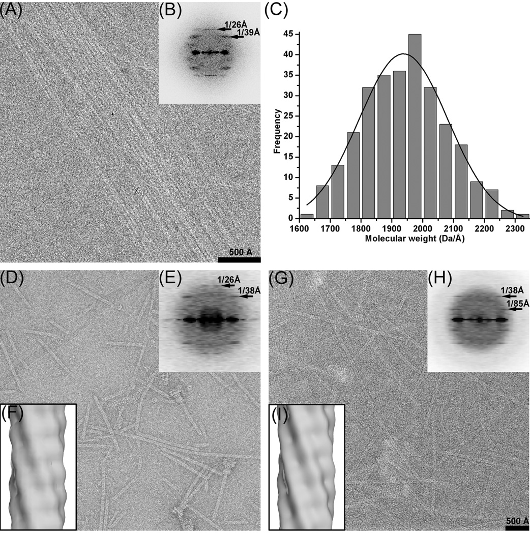
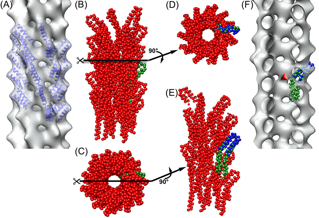
The type III secretion system (T3SS) is essential for the infectivity of many pathogenic Gram-negative bacteria. The T3SS contains proteins that form a channel in the inner and outer bacterial membranes, as well as an extracellular needle that is used for transporting and injecting effector proteins into a host cell. The homology between the T3SS and the bacterial flagellar system has been firmly established, based upon both sequence similarities between respective proteins in the two systems and the structural homology of higher-order assemblies. It has previously been shown that the Shigella flexneri needle has a helical symmetry of approximately 5.6 subunits/turn, which is quite similar to that of the most intensively studied flagellar filament (from Salmonella typhimurium), which has approximately 5.5 subunits/turn. We now show that the Sa. typhimurium needle, expected by homology arguments to be more similar to the Sa. typhimurium flagellar filament than is the needle from Shigella, actually has approximately 6.3 subunits/turn. It is not currently understood how host cell contact, made at the tip of the needle, is communicated to the secretory system at the base. In contrast to the Sa. typhimurium flagellar filament, which shows a nearly crystalline order, the Sa. typhimurium needle has a highly variable symmetry, which could be used to transmit information about host cell contact.
Copyright (c) 2010 Elsevier Ltd. All rights reserved.
Figures
Similar articles
-
The common structural architecture of Shigella flexneri and Salmonella typhimurium type three secretion needles.PLoS Pathog. 2013 Mar;9(3):e1003245. doi: 10.1371/journal.ppat.1003245. Epub 2013 Mar 21. PLoS Pathog. 2013. PMID: 23555258 Free PMC article.
-
Structures of Type III Secretion System Needle Filaments.Curr Top Microbiol Immunol. 2020;427:109-131. doi: 10.1007/82_2019_192. Curr Top Microbiol Immunol. 2020. PMID: 31974760 Review.
-
Helical structure of the needle of the type III secretion system of Shigella flexneri.J Biol Chem. 2003 May 9;278(19):17103-7. doi: 10.1074/jbc.M300091200. Epub 2003 Feb 5. J Biol Chem. 2003. PMID: 12571230
-
Topology and organization of the Salmonella typhimurium type III secretion needle complex components.PLoS Pathog. 2010 Apr 1;6(4):e1000824. doi: 10.1371/journal.ppat.1000824. PLoS Pathog. 2010. PMID: 20368966 Free PMC article.
-
Spinning tails.Curr Opin Struct Biol. 1995 Apr;5(2):187-93. doi: 10.1016/0959-440x(95)80074-3. Curr Opin Struct Biol. 1995. PMID: 7648320 Review.
Cited by
-
Structure and biophysics of type III secretion in bacteria.Biochemistry. 2013 Apr 16;52(15):2508-17. doi: 10.1021/bi400160a. Epub 2013 Apr 5. Biochemistry. 2013. PMID: 23521714 Free PMC article. Review.
-
Research Progress on Small Molecular Inhibitors of the Type 3 Secretion System.Molecules. 2022 Nov 30;27(23):8348. doi: 10.3390/molecules27238348. Molecules. 2022. PMID: 36500441 Free PMC article. Review.
-
Characterization of the interaction between the Salmonella type III secretion system tip protein SipD and the needle protein PrgI by paramagnetic relaxation enhancement.J Biol Chem. 2011 Feb 11;286(6):4922-30. doi: 10.1074/jbc.M110.159434. Epub 2010 Dec 7. J Biol Chem. 2011. PMID: 21138848 Free PMC article.
-
Protein export according to schedule: architecture, assembly, and regulation of type III secretion systems from plant- and animal-pathogenic bacteria.Microbiol Mol Biol Rev. 2012 Jun;76(2):262-310. doi: 10.1128/MMBR.05017-11. Microbiol Mol Biol Rev. 2012. PMID: 22688814 Free PMC article. Review.
-
The common structural architecture of Shigella flexneri and Salmonella typhimurium type three secretion needles.PLoS Pathog. 2013 Mar;9(3):e1003245. doi: 10.1371/journal.ppat.1003245. Epub 2013 Mar 21. PLoS Pathog. 2013. PMID: 23555258 Free PMC article.
References
-
- Galan JE. Salmonella interactions with host cells: type III secretion at work. Annu. Rev. Cell Dev. Biol. 2001;17:53–86. - PubMed
-
- Desvaux M, Hebraud M, Henderson IR, Pallen MJ. Type III secretion: what's in a name? Trends Microbiol. 2006;14:157–160. - PubMed
-
- Cordes FS, et al. Helical structure of the needle of the type III secretion system of Shigella flexneri. J. Biol. Chem. 2003;278:17103–17107. - PubMed
-
- Yonekura K, Maki-Yonekura S, Namba K. Complete atomic model of the bacterial flagellar filament by electron cryomicroscopy. Nature. 2003;424:643–650. - PubMed
Publication types
MeSH terms
Substances
Grants and funding
LinkOut - more resources
Full Text Sources
