

Review
doi: 10.1016/j.ymeth.2010.01.001.
Epub 2010 Jan 12.
Approaches toward super-resolution fluorescence imaging of mitochondrial proteins using PALM
Affiliations
- PMID: 20060907
- PMCID: PMC2938763
- DOI: 10.1016/j.ymeth.2010.01.001
Item in Clipboard
Review
Approaches toward super-resolution fluorescence imaging of mitochondrial proteins using PALM
Methods.
2010 Aug.
Abstract
Mitochondria are difficult targets for microscopy because of their small size and highly compartmentalized, membranous interior. Super-resolution fluorescence microscopy methods have recently been developed that exceed the historical limits of optical imaging. Here we outline considerations and techniques in preparing to image the relative location of mitochondrial proteins using photoactivated localization microscopy (PALM). PALM and similar methods have the capacity to dramatically increase our ability to image proteins within mitochondria, and to expand our knowledge of the location of macromolecules beyond the current limits of immunoEM.
Copyright (c) 2010 Elsevier Inc. All rights reserved.
Figures
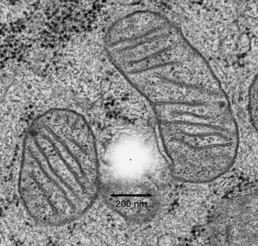
A simulation of the relative space occupied by the diffraction of light from a single fluorescent protein. The white simulated point spread function as might be obtained from a fluorescence microscope is depicted in the center. The point source is represented as a black dot in the middle of the simulated PSF. This is overlaid onto an EM image of mitochondria from NIH3T3 cells. The lateral dimension of the simulated PSF is estimated from a 60X 1.45NA objective lens and is shown with the 200 nm scale bar.
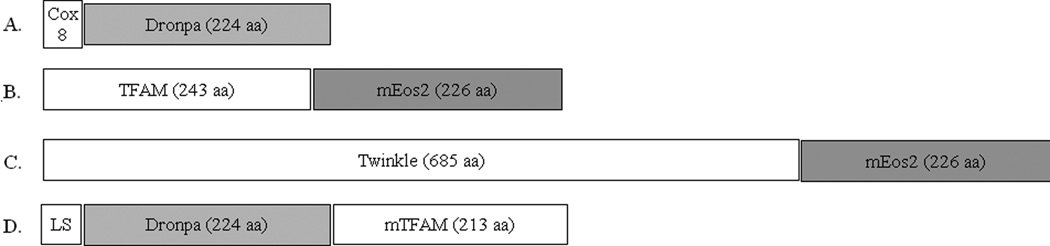
Various mitochondrial fusion protein strategies. A nonfunctional matrix highlighter using only the cleavable mitochondrial localization sequence fused in frame to dronpa is shown in (A). (B) Illustrates a functional TFAM protein fusion with the N-terminus of mEos2. A similar example for the helicase Twinkle is shown in (C) to emphasize the relative scale of fusion proteins. A C-terminal Dronpa fusion is shown in (D), which splits the mature protein sequence from an N-terminal mitochondrial localization sequence (LS). The relative fusion cassettes are to scale based on amino acid sequence length.
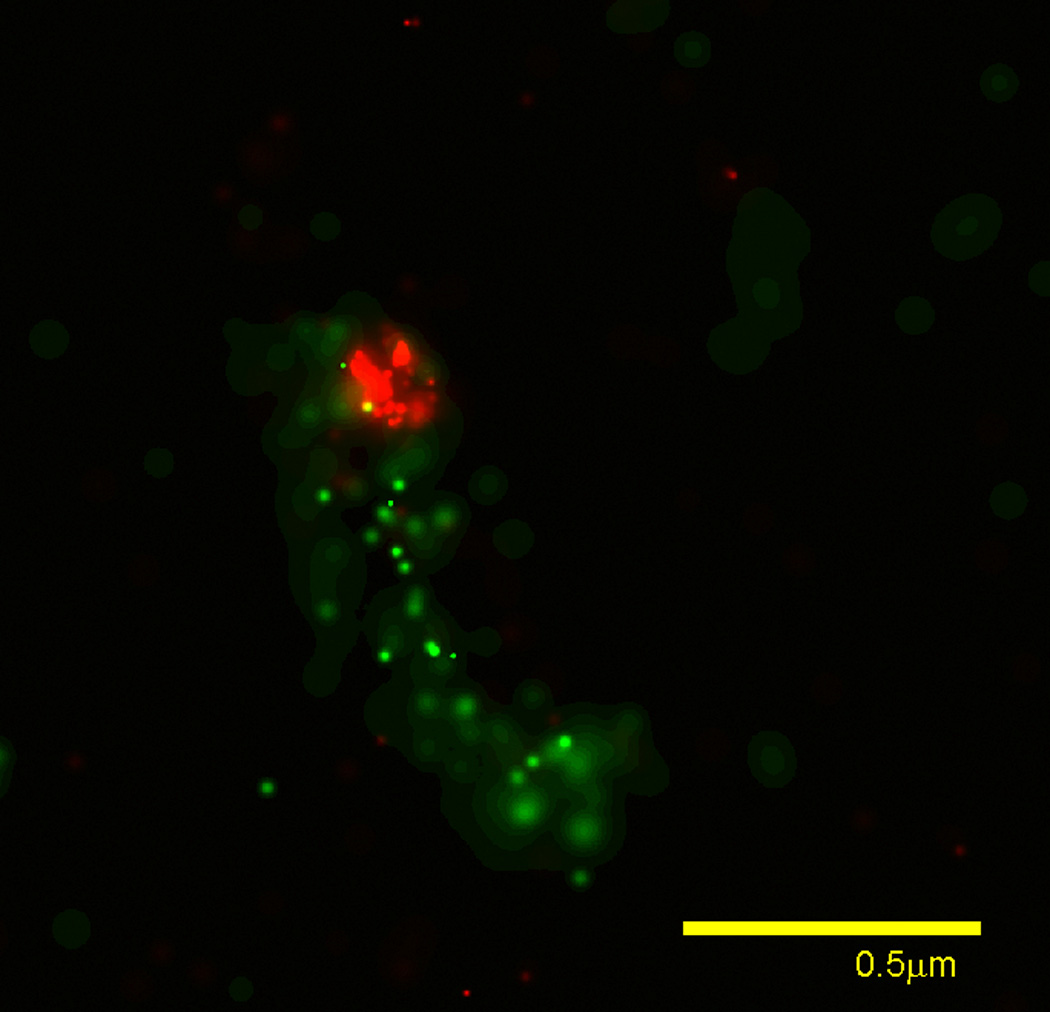
PALM image from an LR-white section. Mitochondrial DNA nucleoid localized TFAM-mEos (red) and matrix localized COX8L-dronpa (green) imaged from a 100 nm thick section of cold polymerized LR-white. Localized molecules are rendered based on confidence of localization as described [5].
Similar articles
-
Correlative photoactivated localization and scanning electron microscopy.PLoS One. 2013 Oct 25;8(10):e77209. doi: 10.1371/journal.pone.0077209. eCollection 2013. PLoS One. 2013. PMID: 24204771 Free PMC article.
-
Monitoring mitophagy in yeast.Methods Enzymol. 2008;451:89-107. doi: 10.1016/S0076-6879(08)03208-4. Methods Enzymol. 2008. PMID: 19185716
-
Superresolution fluorescence imaging of mitochondrial nucleoids reveals their spatial range, limits, and membrane interaction.Mol Cell Biol. 2011 Dec;31(24):4994-5010. doi: 10.1128/MCB.05694-11. Epub 2011 Oct 17. Mol Cell Biol. 2011. PMID: 22006021 Free PMC article.
-
A detailed review of genetically encodable RFPs and far-RFPs and their applications in advanced super-resolution imaging techniques.Biophys Chem. 2025 Jul;322:107432. doi: 10.1016/j.bpc.2025.107432. Epub 2025 Mar 15. Biophys Chem. 2025. PMID: 40117991 Review.
-
Highlights of the optical highlighter fluorescent proteins.J Microsc. 2011 Jul;243(1):1-7. doi: 10.1111/j.1365-2818.2011.03505.x. Epub 2011 May 30. J Microsc. 2011. PMID: 21623791 Free PMC article. Review.
Cited by
-
Mitochondrial ribosomal RNA (rRNA) methyltransferase family members are positioned to modify nascent rRNA in foci near the mitochondrial DNA nucleoid.J Biol Chem. 2013 Oct 25;288(43):31386-99. doi: 10.1074/jbc.M113.515692. Epub 2013 Sep 13. J Biol Chem. 2013. PMID: 24036117 Free PMC article.
-
Continuous organelle separation in an insulator-based dielectrophoretic device.Electrophoresis. 2022 Jun;43(12):1283-1296. doi: 10.1002/elps.202100326. Electrophoresis. 2022. PMID: 34964147 Free PMC article.
-
Correlative photoactivated localization and scanning electron microscopy.PLoS One. 2013 Oct 25;8(10):e77209. doi: 10.1371/journal.pone.0077209. eCollection 2013. PLoS One. 2013. PMID: 24204771 Free PMC article.
-
Insulator-based dielectrophoresis of mitochondria.Biomicrofluidics. 2014 Mar 3;8(2):021801. doi: 10.1063/1.4866852. eCollection 2014 Mar. Biomicrofluidics. 2014. PMID: 24959306 Free PMC article.
-
Scalable Resin Embedding Method for Large-Volume Brain Tissues with High Fluorescence Preservation Capacity.iScience. 2020 Oct 20;23(11):101717. doi: 10.1016/j.isci.2020.101717. eCollection 2020 Nov 20. iScience. 2020. PMID: 33196032 Free PMC article.
References
Publication types
MeSH terms
Substances
Grants and funding
LinkOut - more resources
Full Text Sources
