. 2010 May;170(2):354-63.
doi: 10.1016/j.jsb.2010.01.002. Epub 2010 Jan 11.

Insights into the domain and repeat architecture of target of rapamycin

Affiliations

Insights into the domain and repeat architecture of target of rapamycin

Bruce A Knutson. J Struct Biol. 2010 May.

Abstract

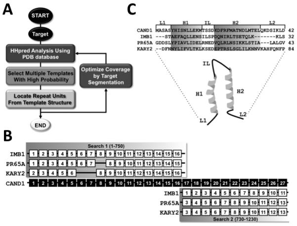
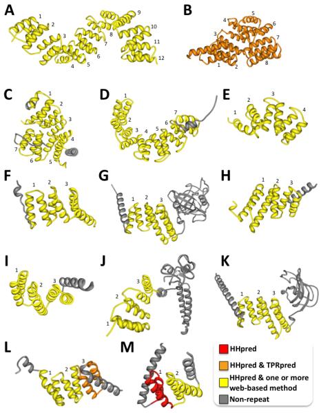
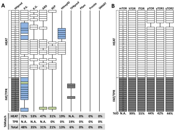
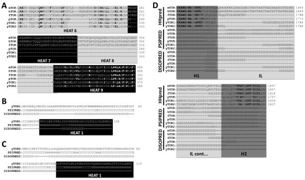
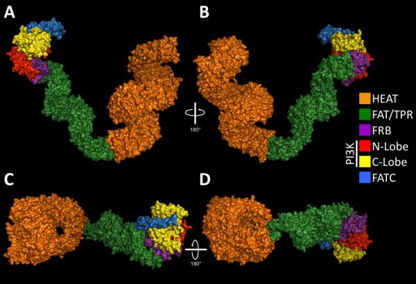
A simple and efficient protein sequence analysis strategy was developed to predict the number and location of structural repeats in the TOR protein. This strategy uses multiple HHpred alignments against proteins of known 3D structure to enable protein repeats referenced from the 3D structure to be traced back to the query protein sequence by using user-directed repeat assignments. The HHpred strategy performed with high sensitivity by predicting 100% of the repeat units within a test set of HEAT- and TPR-repeat-containing proteins of known three-dimensional structure. The HHpred strategy predicts that TOR contains 32 tandem HEAT repeats extending from the N-terminus to the FAT domain, which is itself comprised of 16 tandem TPR repeats. These findings were used to assemble a 3D atomic model for the TOR protein.


Figures

Figure 1
Figure 1. HHpred analysis scheme
A. Flow diagram of the HHpred repeat prediction strategy. B. Identification of individual repeat units from multiple HHpred alignments of the Cand1 target protein and templates of known 3D structure. Black boxes with white numbering represent the number and location of repeat units in the Cand1 protein sequence. Two independent HHpred searches were performed for the Cand1 protein, one with residues 1-750 (Search 1, results shown above) and a second with residues 730-1230 (Search 2, results shown below). HHpred matches the Cand1 protein with high probability to three different template structures that include the importin ß1 (IMB1), protein phosphatase 2A (PR65A), and karyopherin ß2 (KARY2). Each repeat unit was located in the template structures as shown by white boxes and the number of the repeat units within their structure is indicated in black numbering. C. Protein sequence alignments of Cand1 HEAT repeat 1 and template repeat units from IMB1, PR65/A, and KARY2. HEAT repeat structural features are indicated above the sequence alignment and include the following: L1, linker 1, H1, helix 1, IL, interlinker, H2, helix 2, L2, linker 2. The 3D structure of an individual repeat unit is shown below the sequence alignment with the corresponding structural components labeled. H1 and H2 of a single HEAT repeat unit are highlighted in grey and correspond to the grey portions of the ribbon structure while the linkers are drawn in black.
Figure 2
Figure 2. Detection of HEAT repeat units in proteins of known 3D structures
HEAT repeats identified only by HHpred only are colored in red. HEAT repeats identified by HHpred and one or more of the web-based methods are colored in yellow. Grey colored ribbons represent portions of the protein that do not contain HEAT repeat sequences. Individual HEAT repeat units are numbered. Protein structure images were generated by MBT protein workshop available online (http://mbt.sdsc.edu/software/applications) [49]. A. Cand1 (PDB, 1u6g, C), B. CSE1 (PDB, 1wa5, C), C. TNPO1 (PDB, 2z5k, A), D. IMB1 (PDB, 1qgr, A), E. 2AAA (PDB, 1b3u, A), F. AP2α2 (PDB, 2vgl, A), G. CTNB1 (PDB, 1g3j, A), H. VATH (PDB, 1ho8, A), I. IMA1 (PDB, 1bk5, A), J. PKP1 (PDB, 1xm9, A), K. EF3A (PDB, 2iw3, A), L. HSPB1 (PDB, 1xqr, A), M. METH1 (PDB, 1te4, A).
Figure 3
Figure 3. Detection of TPR repeat units in proteins of known 3D structures
TPR repeats identified by HHpred and one or more of the web-based methods are colored in yellow. TPR repeats identified by HHpred and TPRpred only are colored in orange while TPR repeats identified only by HHpred are colored in red. Grey colored ribbons represent portions of the protein that do not contain HEAT repeat sequences. Individual TPR repeat units are numbered. Protein structure images were generated by MBT protein workshop available online (http://mbt.sdsc.edu/software/applications) [49]. A. OGT1 (PDB, 1w3b, A), B. MALT (PDB, 1hz4, A), C. PEX5 (PDB, 1fch, A), D. HCPC (PDB, 1ouv, A), E. HCPB (PDB, 1klx, A), F. PPP5 (PDB, 1wao, A), G. PPID (PDB, 1ihg, A), H. PLAF7 (PDB, 2fbn, A), I. STIP1 (PDB, 1elr, A), J. STUB1 (PDB, 2c2l, A), K. FKBP4 (PDB, 1qz2, A), L. SYCD (PDB, 2vgx, A), M. FIS1 (PDB, 1nzn, A).
Figure 4
Figure 4. Prediction and comparison of TOR HEAT and TPR repeats
Rectangular boxes denote individual repeat units. A solid black line represents a gap in the structure corresponding to a missing repeat unit relative to the HHpred predicted structure. Individual repeat units in the mTOR structure are numbered within each rectangular box. A. The method used to predict the protein repeats is indicated at the top of each column. P.K., Perry and Klechner prediction. K.G., Kippert and Gerloff protocol. White and grey colored boxes indicate a HEAT and TPR repeats correctly identified by both HHpred and web-based method, respectively. Blue boxes indicate a mismatch between the HHpred and web-based method. Green boxes indicate a HEAT repeat identified by a web-based method that matches with a TPR repeat identified by HHpred. B. The HEAT and FAT/TPR domains from six different TOR proteins are shown. The name of each TOR protein is labeled at the top of each structure. Percent identity of each protein relative to the mTOR protein sequence is indicated at the bottom of the structures. White and grey rectangular boxes denote HEAT and TPR repeat units, respectively
Figure 5
Figure 5. Sequence analysis of divergent TOR repeats structures
Panel A shows a multiple sequence alignment of HEAT 6-9 for six TOR protein sequences from different eukaryotic species. The name of each TOR protein is indicated at the left of each sequence. Grey and black boxes indicate individual HEAT repeat units that are labeled at the bottom of each box. The numbers to the right of the sequence denote amino acid position. Bold lettering indicates an amino acid conserved for at least four of the sequences. Protein sequence analysis of yTOR1 (Panel B) and yTOR2 (Panel C) N-terminal extensions by PSIPRED (middle row) and DISOPRED2 (bottom row). H, helix, E, beta sheet, C, coil, D, disordered, =, ordered, −, gap. The black box and white lettering indicates predicted HEAT repeat 1 of yTOR1 and 2. Panel D shows a multiple sequence alignment and predicted secondary structure of TPR repeat 13. The first row labeled HHpred depicts the HMM alignments generated by HHpred that were combined for six different TOR protein sequences. Below the primary structure alignment, the secondary structures predicted by HHpred using PSIPRED are listed for each TOR protein amino acid position. The final row labeled DISOPRED2 shows disordered protein sequence predictions that are listed for each TOR protein amino acid position. D, disordered, =, ordered. Dark and light grey boxes indicate the two helices and interlinker of TPR repeat 13, respectively. H1, Helix 1, H2, Helix 2, IL, interlinker.
Figure 6
Figure 6. 3D model of mTOR based on HHpred results
Panels A-D show different views of the TOR 3D model built using the HHpred results listed in supplementary Table 3. Structures were corrected manually to minimize insertions of the template structure that are not found in the target protein. Panels A and B show side views while Panels C and D show top and bottom views, respectively. The 3D model was built in pymol. Orange and green colors correspond to HEAT and TPR/FAT domains, respectively. Purple and blue colors correspond to the FRB and FATC domains, respectively. Yellow and Red corresponds to the N- and C-lobe of the PI3K catalytic domain, respectively.

Similar articles

Cited by

References

    1. Chen J, et al. Identification of an 11-kDa FKBP12-rapamycin-binding domain within the 289-kDa FKBP12-rapamycin-associated protein and characterization of a critical serine residue. Proc Natl Acad Sci U S A. 1995;92(11):4947–51. - PMC - PubMed
    1. Choi J, et al. Structure of the FKBP12-rapamycin complex interacting with the binding domain of human FRAP. Science. 1996;273(5272):239–42. - PubMed
    1. Heitman J, Movva NR, Hall MN. Targets for cell cycle arrest by the immunosuppressant rapamycin in yeast. Science. 1991;253(5022):905–9. - PubMed
    1. Helliwell SB, et al. TOR1 and TOR2 are structurally and functionally similar but not identical phosphatidylinositol kinase homologues in yeast. Mol Biol Cell. 1994;5(1):105–18. - PMC - PubMed
    1. Kunz J, et al. Target of rapamycin in yeast, TOR2, is an essential phosphatidylinositol kinase homolog required for G1 progression. Cell. 1993;73(3):585–96. - PubMed

Publication types

MeSH terms

Substances