

Antibacterial action of a novel functionalized chitosan-arginine against Gram-negative bacteria
- PMID: 20060936
- PMCID: PMC2874111
- DOI: 10.1016/j.actbio.2010.01.002
Antibacterial action of a novel functionalized chitosan-arginine against Gram-negative bacteria
Abstract
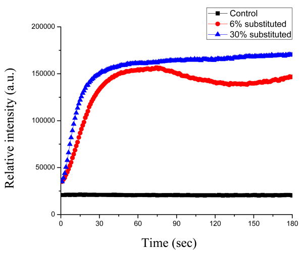
The antimicrobial activity of chitosan and chitosan derivatives has been well established. However, although several mechanisms have been proposed, the exact mode of action is still unclear. Here we report on the investigation of antibacterial activity and the antibacterial mode of action of a novel water-soluble chitosan derivative, arginine-functionalized chitosan, on the Gram-negative bacteria Pseudomonas fluorescens and Escherichia coli. Two different arginine-functionalized chitosans (6% arginine-substituted and 30% arginine-substituted) each strongly inhibited P. fluorescens and E. coli growth. Time-dependent killing efficacy experiments showed that 5000 mg l(-1) of 6%- and 30%-substituted chitosan-arginine killed 2.7 logs and 4.5 logs of P. fluorescens, and 4.8 logs and 4.6 logs of E. coli in 4h, respectively. At low concentrations, the 6%-substituted chitosan-arginine was more effective in inhibiting cell growth even though the 30%-substituted chitosan-arginine appeared to be more effective in permeabilizing the cell membranes of both P. fluorescens and E. coli. Studies using fluorescent probes, 1-N-phenyl-naphthylamine (NPN), nile red (NR) and propidium iodide (PI), and field emission scanning electron microscopy (FESEM) suggest that chitosan-arginine's antibacterial activity is, at least in part, due to its interaction with the cell membrane, in which it increases membrane permeability.
Copyright 2010 Acta Materialia Inc. Published by Elsevier Ltd. All rights reserved.
Figures


Similar articles
-
Antibacterial activity of ozone-depolymerized crawfish chitosan.J Food Sci. 2008 Oct;73(8):M400-4. doi: 10.1111/j.1750-3841.2008.00922.x. J Food Sci. 2008. PMID: 19019121
-
Antimicrobial effect and mechanism of non-antibiotic alkyl gallates against Pseudomonas fluorescens on the surface of Russian sturgeon (Acipenser gueldenstaedti).Int J Food Microbiol. 2021 Mar 16;342:109093. doi: 10.1016/j.ijfoodmicro.2021.109093. Epub 2021 Feb 11. Int J Food Microbiol. 2021. PMID: 33607540
-
Antibacterial activity of chemically defined chitosans: influence of molecular weight, degree of acetylation and test organism.Int J Food Microbiol. 2011 Jul 15;148(1):48-54. doi: 10.1016/j.ijfoodmicro.2011.04.023. Epub 2011 Apr 30. Int J Food Microbiol. 2011. PMID: 21605923
-
Gallic acid-grafted-chitosan inhibits foodborne pathogens by a membrane damage mechanism.J Agric Food Chem. 2013 Jul 3;61(26):6574-9. doi: 10.1021/jf401254g. Epub 2013 May 14. J Agric Food Chem. 2013. PMID: 23635088
-
Preparation, characterization and antibacterial properties of 6-deoxy-6-arginine modified chitosan.Carbohydr Polym. 2020 Feb 15;230:115635. doi: 10.1016/j.carbpol.2019.115635. Epub 2019 Nov 21. Carbohydr Polym. 2020. PMID: 31887858
Cited by
-
Designing polysaccharide-based antibacterial biomaterials for wound healing applications.Biomatter. 2011 Oct-Dec;1(2):189-97. doi: 10.4161/biom.19005. Biomatter. 2011. PMID: 23507748 Free PMC article.
-
Nanoarchitectonics for Biodegradable Superabsorbent Based on Carboxymethyl Starch and Chitosan Cross-Linked with Vanillin.Int J Mol Sci. 2022 May 11;23(10):5386. doi: 10.3390/ijms23105386. Int J Mol Sci. 2022. PMID: 35628197 Free PMC article.
-
Multi-Target Antibacterial Mechanism of Moringin From Moringa oleifera Seeds Against Listeria monocytogenes.Front Microbiol. 2022 Jun 8;13:925291. doi: 10.3389/fmicb.2022.925291. eCollection 2022. Front Microbiol. 2022. PMID: 35756047 Free PMC article.
-
Antibacterial activity of lysozyme-chitosan oligosaccharide conjugates (LYZOX) against Pseudomonas aeruginosa, Acinetobacter baumannii and Methicillin-resistant Staphylococcus aureus.PLoS One. 2019 May 28;14(5):e0217504. doi: 10.1371/journal.pone.0217504. eCollection 2019. PLoS One. 2019. PMID: 31136634 Free PMC article.
-
Photo-Crosslinked Polymeric Matrix with Antimicrobial Functions for Excisional Wound Healing in Mice.Nanomaterials (Basel). 2018 Oct 5;8(10):791. doi: 10.3390/nano8100791. Nanomaterials (Basel). 2018. PMID: 30301173 Free PMC article.
References
-
- Lim SH, Hudson SM. Review of chitosan and its derivatives as antimicrobial agents and their uses as textile chemicals. J Macromol Sci Part C – Polymer Reviews. 2003;C43:223–269.
-
- Didenko LV, Gerasimenko DV, Konstantinova ND, Silkina TA, Avdienko ID, Bannikova GE, et al. Ultrastructural study of chitosan effects on Klebsiella and Staphylococci. Bull Exp Biol Med. 2005;140:343–347. - PubMed
-
- Helander IM, Nurmiaho-Lassila EL, Ahvenainen R, Rhoades J, Roller S. Chitosan disrupts the barrier properties of the outer membrane of gram-negative bacteria. Int J Food Microbiol. 2001;71:235–244. - PubMed
-
- Bae K, Jun EJ, Lee SM, Paik DI, Kim JB. Effect of water-soluble reduced chitosan on Streptococcus mutans, plaque regrowth and biofilm vitality. Clin Oral Invest. 2006;10:102–107. - PubMed
Publication types
MeSH terms
Substances
Grants and funding
LinkOut - more resources
Full Text Sources
Other Literature Sources
Medical
Research Materials
Miscellaneous
