

Human eyelid meibomian glands and tarsal muscle are recognized by autoantibodies from patients affected by a new variant of endemic pemphigus foliaceus in El-Bagre, Colombia, South America
- PMID: 20061054
- PMCID: PMC2992456
- DOI: 10.1016/j.jaad.2009.06.007
Human eyelid meibomian glands and tarsal muscle are recognized by autoantibodies from patients affected by a new variant of endemic pemphigus foliaceus in El-Bagre, Colombia, South America
Abstract
Background: Previously, we described a new variant of endemic pemphigus foliaceus (EPF) in Colombia, South America (El Bagre-EPF).
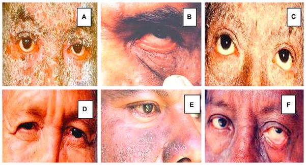
Objective: Continuing our characterization of this variant of EPF, we now focus on one of our previously reported clinical findings: the presence of ocular lesions. These ocular lesions are seen in patients having extensive skin involvement, as measured by the Lund and Browder scale, which is generally used for patients with skin burns.
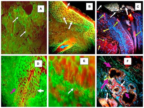
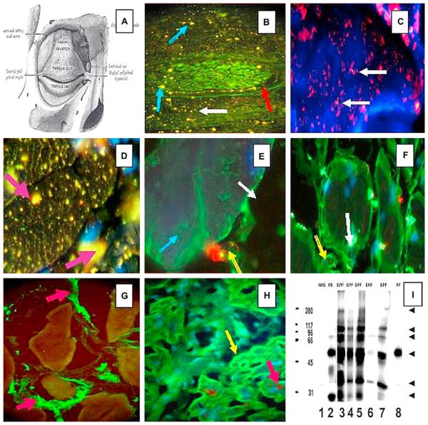
Methods: We specifically searched for evidence of autoreactivity to various eyelid structures in these patients and correlated our immunologic data with the clinical findings. We performed indirect immunofluorescence studies using normal-appearing human eyelid skin from routine blepharoplasties as substrate tissue. We tested sera from 12 patients with El Bagre-EPF and ocular lesions, 5 patients with sporadic (nonendemic) pemphigus foliaceus, and 20 healthy control subjects (10 from the El Bagre-EPF endemic area and 10 from nonendemic areas). We used fluorescein isothiocyanate conjugated goat antiserum to human total IgG/IgA/IgM as a secondary antibody. In addition, we used fluorescein isothiocyanate conjugated antibodies to human fibrinogen, albumin, IgG, IgE, C1q, and C3, Texas Red (Rockland Immunochemicals, Inc, Gilbertsville, PA), Alexa Fluor 555, or Alexa Fluor 594 (Invitrogen, Carlsbad, CA). Ki-67 (a cell proliferation marker) was used to determine the cell proliferation rate, and nuclear counterstaining was performed with either 4', 6-diamidino-2-phenylindole or Topro III (Invitrogen, Carlsbad, CA).
Results: We observed autoreactivity to multiple eyelid structures, including meibomian glands and tarsal muscle bundles at different levels, and some areas of the epidermis and the dermis close to the isthmus of the eyelids. Tarsal plate autoreactivity was seen in 10 of 12 of the El Bagre-EPF sera and in one control with pemphigus erythematosus. Furthermore, immunoprecipitation using an eyelid sample as a substrate with 1 mmol/L of sodium orthovanodate showed autoreactivity to several antigens, including some of possible lipid origin.
Limitations: The main limitation of this study is the fact that the antigen or antigens remain unknown.
Conclusion: We identified for the first time to our knowledge autoantibodies to meibomian glands and tarsal muscle in El Bagre-EPF. Our findings suggest that the autoantibodies to the ocular structures cause the clinical and histopathological findings in the ocular lesions in El Bagre-EPF.
Copyright 2009 American Academy of Dermatology, Inc. Published by Mosby, Inc. All rights reserved.
Figures
Similar articles
-
Antibodies to pilosebaceous units along their neurovascular supply routes in a new variant of endemic pemphigus foliaceus in Colombia, South America.Eur J Dermatol. 2011 May-Jun;21(3):371-5. doi: 10.1684/ejd.2011.1310. Eur J Dermatol. 2011. PMID: 21543287
-
Patients with a new variant of endemic pemphigus foliaceus have autoantibodies against arrector pili muscle, colocalizing with MYZAP, p0071, desmoplakins 1 and 2 and ARVCF.Clin Exp Dermatol. 2017 Dec;42(8):874-880. doi: 10.1111/ced.13214. Epub 2017 Oct 15. Clin Exp Dermatol. 2017. PMID: 29034528
-
Palm tissue displaying a polyclonal autoimmune response in patients affected by a new variant of endemic pemphigus foliaceus in Colombia, South America.Eur J Dermatol. 2010 Jan-Feb;20(1):74-81. doi: 10.1684/ejd.2010.0834. Epub 2009 Nov 4. Eur J Dermatol. 2010. PMID: 19889592
-
Rare clinical form in two patients affected by a new variant of endemic pemphigus in northern Colombia.Skinmed. 2004 Nov-Dec;3(6):317-21. doi: 10.1111/j.1540-9740.2004.03514.x. Skinmed. 2004. PMID: 15538080 Review.
-
Brazilian pemphigus foliaceus, endemic pemphigus foliaceus, or fogo selvagem (wild fire).Dermatol Clin. 1994 Oct;12(4):765-76. Dermatol Clin. 1994. PMID: 7805306 Review.
Cited by
-
Markers for Sebaceoma Show a Spectrum of Cell Cycle Regulators, Tumor Suppressor Genes, and Oncogenes.N Am J Med Sci. 2015 Jun;7(6):275-80. doi: 10.4103/1947-2714.159338. N Am J Med Sci. 2015. PMID: 26199925 Free PMC article.
-
Overexpression of linker for activated T cells, cyclooxygenase-2, CD1a, CD68 and myeloid/histiocyte antigen in an inflamed seborrheic keratosis.N Am J Med Sci. 2011 Mar;3(3):161-3. doi: 10.4297/najms.2011.3161. N Am J Med Sci. 2011. PMID: 22540084 Free PMC article.
-
Endemic pemphigus over a century: Part II.N Am J Med Sci. 2010 Mar;2(3):114-25. N Am J Med Sci. 2010. PMID: 22624125 Free PMC article.
-
Cell junction protein armadillo repeat gene deleted in velo-cardio-facial syndrome is expressed in the skin and colocalizes with autoantibodies of patients affected by a new variant of endemic pemphigus foliaceus in Colombia.Dermatol Pract Concept. 2017 Oct 31;7(4):3-8. doi: 10.5826/dpc.0704a02. eCollection 2017 Oct. Dermatol Pract Concept. 2017. PMID: 29214101 Free PMC article.
-
An inflamed trichilemmal (pilar) cyst: Not so simple?N Am J Med Sci. 2011 Sep;3(9):431-4. doi: 10.4297/najms.2011.3431. N Am J Med Sci. 2011. PMID: 22362454 Free PMC article.
References
-
- Viera J. Pemphigus foliaceus (fogo selvagem): endemic disease of Sao Paolo (Brazil) Arch Dermatol Syph. 1940;211:858.
-
- Proenca N, Ribeira A. Aspectus epidemiologicos do penfigo foliaceo no Brazil [Epidemiologic features of pemphigus foliaceus in Brazil] Rev Assoc Med Bras. 1976;22:281–4. - PubMed
-
- Castro R, Proenca N. Semelhancas e diferencas entre o fogo selvagem e o penfigo foliaceo de Cazenave [Similarities and differences between South American pemphigus foliaceus and Cazanave's pemphigus foliaceus] An Bras Dermatol. 1983;53:137–9.
-
- Diaz L, Sampaio S, Rivitti EA, Martins CR, Cunha PR, Lombardi C, et al. Endemic pemphigus foliaceus (fogo selvagem): clinical features and immunopathology. J Am Acad Dermatol. 1989;20:657–9. - PubMed
-
- Abréu-Vélez AM, Beutner EH, Montoya F, Bollag WB, Hashimoto T. Analyses of autoantigens in a new form of endemic pemphigus foliaceus in Colombia. J Am Acad Dermatol. 2003;49:609–14. - PubMed
Publication types
MeSH terms
Substances
Grants and funding
LinkOut - more resources
Full Text Sources
Medical
Miscellaneous
