Clinical Trial
. 2010 Apr;99(4):333-45.
doi: 10.1016/j.ymgme.2009.12.007. Epub 2010 Jan 12.

Classical maple syrup urine disease and brain development: principles of management and formula design

Affiliations
Clinical Trial

Classical maple syrup urine disease and brain development: principles of management and formula design

Kevin A Strauss et al. Mol Genet Metab. 2010 Apr.

Erratum in

  • Mol Genet Metab. 2011 Jun;103(2):202. Shelmer, Diana [corrected to Shellmer, Diana]

Abstract

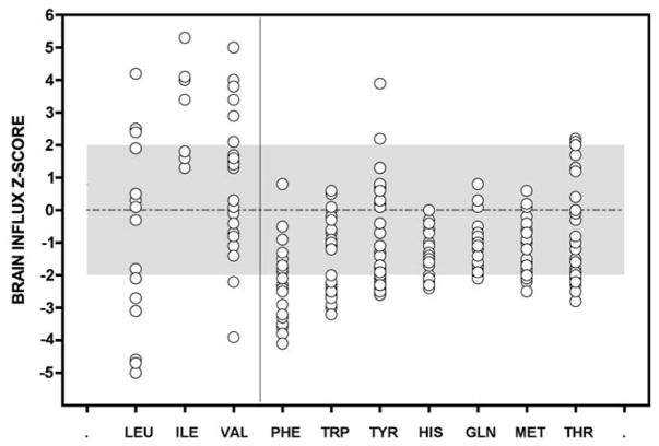
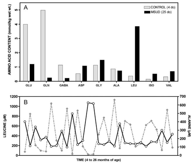
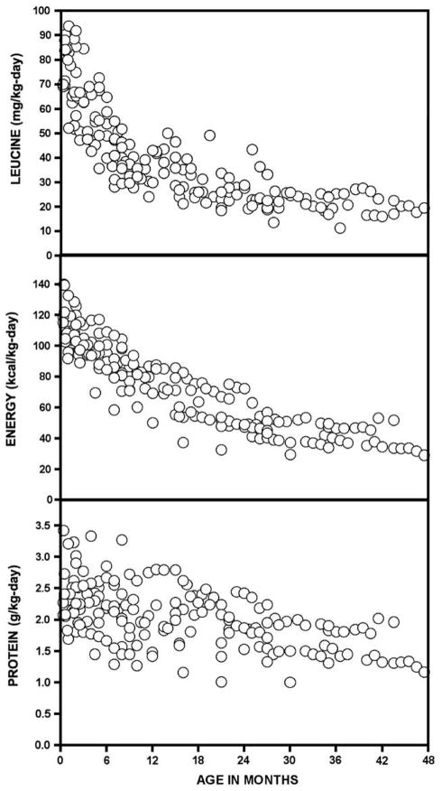
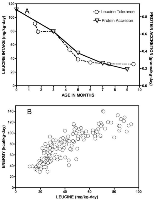
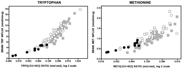
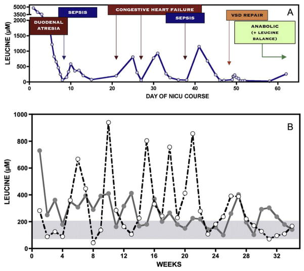
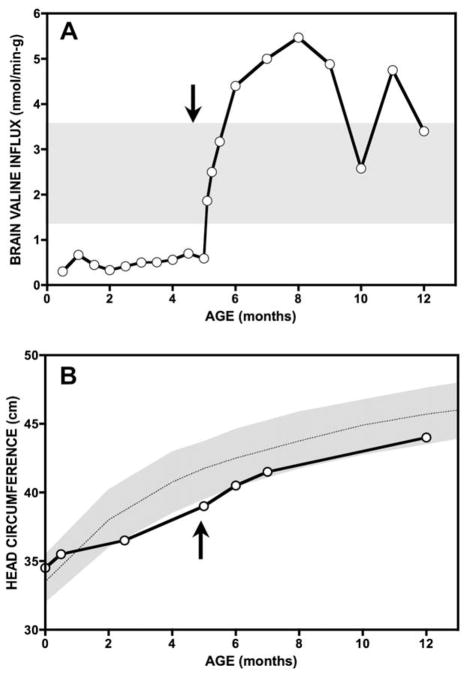
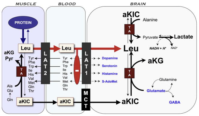
Branched-chain ketoacid dehydrogenase deficiency results in complex and volatile metabolic derangements that threaten brain development. Treatment for classical maple syrup urine disease (MSUD) should address this underlying physiology while also protecting children from nutrient deficiencies. Based on a 20-year experience managing 79 patients, we designed a study formula to (1) optimize transport of seven amino acids (Tyr, Trp, His, Met, Thr, Gln, Phe) that compete with branched-chain amino acids (BCAAs) for entry into the brain via a common transporter (LAT1), (2) compensate for episodic depletions of glutamine, glutamate, and alanine caused by reverse transamination, and (3) correct deficiencies of omega-3 essential fatty acids, zinc, and selenium widespread among MSUD patients. The formula was enriched with LAT1 amino acid substrates, glutamine, alanine, zinc, selenium, and alpha-linolenic acid (18:3n-3). Fifteen Old Order Mennonite children were started on study formula between birth and 34 months of age and seen at least monthly in the office. Amino acid levels were checked once weekly and more often during illnesses. All children grew and developed normally over a period of 14-33 months. Energy demand, leucine tolerance, and protein accretion were tightly linked during periods of normal growth. Rapid shifts to net protein degradation occurred during illnesses. At baseline, most LAT1 substrates varied inversely with plasma leucine, and their calculated rates of brain uptake were 20-68% below normal. Treatment with study formula increased plasma concentrations of LAT1 substrates and normalized their calculated uptakes into the nervous system. Red cell membrane omega-3 polyunsaturated fatty acids and serum zinc and selenium levels increased on study formula. However, selenium and docosahexaenoic acid (22:6n-3) levels remained below normal. During the study period, hospitalizations decreased from 0.35 to 0.14 per patient per year. There were 28 hospitalizations managed with MSUD hyperalimentation solution; 86% were precipitated by common infections, especially vomiting and gastroenteritis. The large majority of catabolic illnesses were managed successfully at home using 'sick-day' formula and frequent amino acid monitoring. We conclude that the study formula is safe and effective for the treatment of classical MSUD. In principle, dietary enrichment protects the brain against deficiency of amino acids used for protein accretion, neurotransmitter synthesis, and methyl group transfer. Although the pathophysiology of MSUD can be addressed through rational formula design, this does not replace the need for vigilant clinical monitoring, frequent measurement of the complete amino acid profile, and ongoing dietary adjustments that match nutritional intake to the metabolic demands of growth and illness.


Figures

Fig. 1
Fig. 1
Uptake values of 10 amino acids competing for a common transporter (LAT1) at the blood–brain barrier were calculated from plasma amino acid profiles of a Mennonite boy drinking a standard infant MSUD formula. Samples were collected during the 22 months before the trial period. Uptake values are expressed as z-scores, where z = [(patient value − control mean)/control standard deviation]. Normal values, taken from 52 control subjects, lie between +2 and −2 (gray area). Averaged over long periods, brain uptake of most LAT1 substrates is low (right panel). Uptake values for BCAAs (left panel) are highly variable, reflecting the unstable BCAA homeostasis characteristic of classical MSUD.
Fig. 2
Fig. 2
In brain, aKIC likely depletes glutamate through reverse transamination to leucine and alpha-ketoglutarate; this in turn affects brain content of GABA and glutamine (See Fig. 8). (A) In 1966, Prensky and Moser compared brain free amino acids from a 25 day-old infant who died in MSUD crisis (black bars) to a 4 day-old infant who did not have the disease (gray bars). They found profound depletion of glutamate, glutamine, and GABA in MSUD brain tissue (Ref. [15]). A similar phenomenon is observed in cultured astrocytes, Dbt−/− mice, and Poll-Hereford calves with MSUD (Refs. [–14]). (B) The predominant effect of elevated aKIC in muscle may be to deplete alanine to form leucine and pyruvate. In a Mennonite child, plasma leucine (solid line) and alanine (dashed line) are reciprocally related over a 22-month period (rs = −0.86, p < 0.0001), suggesting this depletion mechanism operates in vivo (See Table 1).
Fig. 3
Fig. 3
Serial leucine, energy, and protein intakes of 15 Mennonite infants with MSUD treated with study formula.
Fig. 4
Fig. 4
(A) In infants with classical MSUD, BCKDH activity is absent and urine and insensible losses of leucine are negligible. Thus, leucine tolerance naturally traces the balance between protein degradation and synthesis. (B) Protein accretion is energetically costly, illustrated by the close correlation between leucine tolerance (x-axis) and energy demand (y-axis) during the first 4 years of life (rs = 0.9, p < 0.0001).
Fig. 5
Fig. 5
The panels depict how the concentration ratio of an LAT1 substrate (e.g., tryptophan or methionine) to the sum of leucine and isoleucine (x-axis) relates to its calculated uptake across the blood–brain barrier (y-axis). Before the formula trial, low concentration ratios were associated with low brain uptake (black circles). Treatment with study formula (white circles) reduced leucine and isoleucine levels while simultaneously increasing plasma concentrations of other LAT1 substrates (See Table 5). Incrased competitor: [leucine + isoleucine] ratios increased calculated brain uptakes to the control range (gray squares; N = 52). Note the x-axis is a log 2 scale.
Fig. 6
Fig. 6
(A) Over a 62-day NICU course, a Mennonite infant with Down syndrome suffered from duodenal atresia, large ventricular septal defect, and two episodes of sepsis. Serial monitoring shows the affect of catabolic stress on plasma leucine levels, in each case treated with parenteral nutritional therapy to restore net protein anabolism. (B) The figure depicts once weekly plasma leucine values over a 34-week period from two infants, one shown with a solid line and the other with a dashed line. Leucine levels are erratic relative to the normal homeostatic range (blue shaded area), and metabolic control varies considerably between individuals. Episodic leucine elevations (white circles) reflect transient catabolic states triggered by common infections (e.g., gastroenteritis, otitis, bronchiolitis, etc.).
Fig. 7
Fig. 7
A 5 month-old infant from the Netherlands presented with growth failure, microcephaly, skin desquamation, and normocytic anemia. Plasma valine levels between 30 and 90 μM over preceding months predicted low valine uptake into the nervous system. Aggressive valine supplementation to maintain plasma levels at least twice the leucine concentration were associated with a large calculated increase of tissue valine uptake (A), skin healing, resolution of anemia, and a surge in brain growth and myelination (B).
Fig. 8
Fig. 8
Pathophysiology of brain disease in MSUD. Episodic elevations of BCAAs occur when muscle degrades protein in response to infection or other physiologic stress and releases BCAAs and their ketoacid derivatives. High aKIC in muscle can reverse flow through cytosolic transaminases and deplete tissue of alanine and other amino acids. As leucine (Leu) exits tissue via the large amino acid transporter type 2 (LAT2), it drives import of other amino acids (heteroexchange). This process increases the relative concentration of leucine to other amino acids in plasma. At the blood–brain barrier, leucine, which has a low Km for LAT1, saturates the transporter and blocks uptake of its competitors, including precursors for neurotransmitters (dopamine, norepinephrine, serotonin, and histamine) and S-adenosylmethionine (S-AdoMet), the brain’s major methyl donor. Alpha-ketoisocaproic acid (aKIC) enters the brain via the monocarboxylate transporter (MCT) and reverses flux through cerebral transaminases (TA). This depletes brain glutamate, GABA, and glutamine while increasing production of leucine and alpha-ketoglutarate (aKG). Glutamate and GABA are the most abundant excitatory and inhibitory neurotransmitters, respectively, in the human brain. MSUD encephalopathy may also block oxidative phosphorylation through an as yet unknown mechanism; the increased NADH/NAD ratio could explain high cerebral lactate levels observed in both mice and humans during metabolic crisis.

Similar articles

Cited by

References

    1. Strauss KA, Morton DH. Branched-chain ketoacyl dehydrogenase deficiency: maple syrup disease. Curr Treat Options Neurol. 2003;5:329–341. - PubMed
    1. Morton DH, Morton CS, Strauss KA, Robinson DL, Puffenberger EG, Hendrickson C, Kelley RI. Pediatric medicine and the genetic disorders of the Amish and Mennonite people of Pennsylvania. Am J Med Genet C Semin Med Genet. 2003;121:5–17. - PubMed
    1. Strauss KA, Puffenberger EG, Morton DH. GeneReviews. University of Washington; Seattle: 2006. Maple Syrup Urine Disease. - PubMed
    1. Kamei A, Takashima S, Chan F, Becker LE. Abnormal dendritic development in maple syrup urine disease. Pediatr Neurol. 1992;8:145–147. - PubMed
    1. Smith QR, Stoll JS. Blood–brain barrier amino acid transport. In: Pardridge WM, editor. Introduction to the Blood–Brain Barrier. Cambridge University Press; Cambridge: 1998. pp. 188–197.

Publication types