. 2010 Feb;9(2):149-58.
doi: 10.1016/S1474-4422(10)70002-8. Epub 2010 Jan 8.

Differential diagnosis of parkinsonism: a metabolic imaging study using pattern analysis

Affiliations

Differential diagnosis of parkinsonism: a metabolic imaging study using pattern analysis

Chris C Tang et al. Lancet Neurol. 2010 Feb.

Abstract

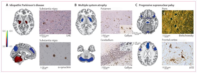
Background: Idiopathic Parkinson's disease can present with symptoms similar to those of multiple system atrophy or progressive supranuclear palsy. We aimed to assess whether metabolic brain imaging combined with spatial covariance analysis could accurately discriminate patients with parkinsonism who had different underlying disorders.

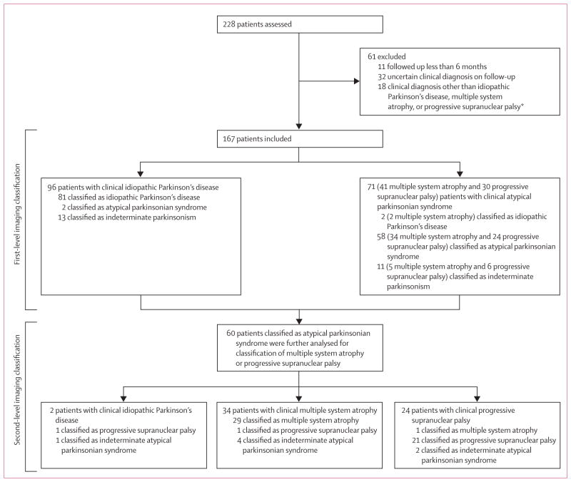
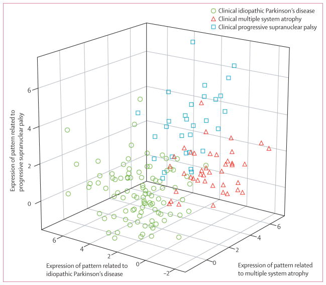
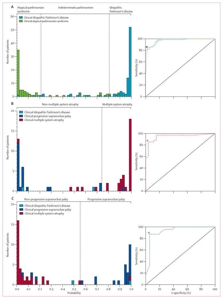
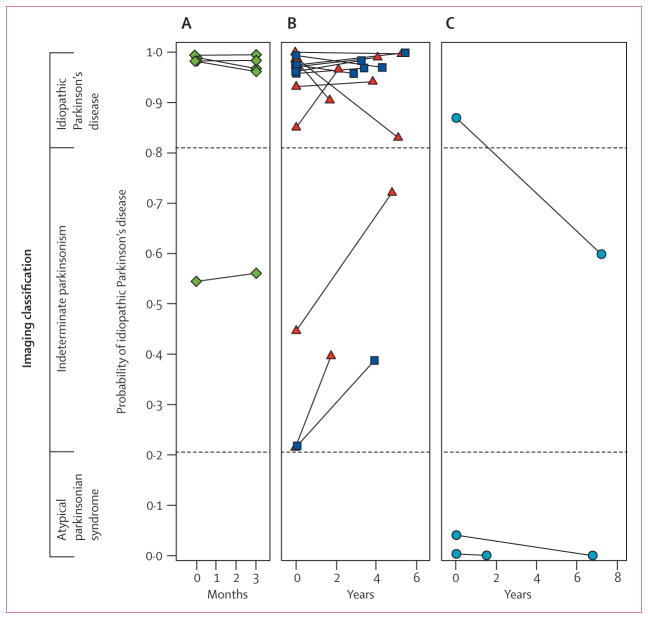
Methods: Between January, 1998, and December, 2006, patients from the New York area who had parkinsonian features but uncertain clinical diagnosis had fluorine-18-labelled-fluorodeoxyglucose-PET at The Feinstein Institute for Medical Research. We developed an automated image-based classification procedure to differentiate individual patients with idiopathic Parkinson's disease, multiple system atrophy, and progressive supranuclear palsy. For each patient, the likelihood of having each of the three diseases was calculated by use of multiple disease-related patterns with logistic regression and leave-one-out cross-validation. Each patient was classified according to criteria defined by receiver-operating-characteristic analysis. After imaging, patients were assessed by blinded movement disorders specialists for a mean of 2.6 years before a final clinical diagnosis was made. The accuracy of the initial image-based classification was assessed by comparison with the final clinical diagnosis.

Findings: 167 patients were assessed. Image-based classification for idiopathic Parkinson's disease had 84% sensitivity, 97% specificity, 98% positive predictive value (PPV), and 82% negative predictive value (NPV). Imaging classifications were also accurate for multiple system atrophy (85% sensitivity, 96% specificity, 97% PPV, and 83% NPV) and progressive supranuclear palsy (88% sensitivity, 94% specificity, 91% PPV, and 92% NPV).

Interpretation: Automated image-based classification has high specificity in distinguishing between parkinsonian disorders and could help in selecting treatment for early-stage patients and identifying participants for clinical trials.

Funding: National Institutes of Health and General Clinical Research Center at The Feinstein Institute for Medical Research.


Conflict of interest statement

Conflicts of interest

DE is co-inventor of US patents 5 632 276 (filed Jan 27, 1995, granted May 27, 1997) and 5 873 823 (filed Sept 4, 1996, granted Feb 23, 1999) for the use of spatial patterns for the diagnosis of brain disease. DE has no financial conflicts of interest. All other authors have no conflicts of interest.

Figures

Figure 1
Figure 1. Study protocol
*This group included seven patients with corticobasal ganglionic degeneration, three with infectious parkinsonism or prion disease, three with vascular parkinsonism with MRI abnormalities, one with psychogenic disease, one with an autoimmune disorder, one with rapid-onset dystonia-parkinsonism, one with progressive lateral sclerosis with atypical parkinsonism, and one with multiple sclerosis and idiopathic Parkinson’s disease.
Figure 2
Figure 2
3D plot of FDG-PET pattern expression
Figure 3
Figure 3. Predicted disease probability for differential diagnosis of parkinsonism
Frequency distributions (left) for idiopathic Parkinson’s disease and atypical parkinsonian syndrome (A), multiple system atrophy (B), and progressive supranuclear palsy (C). The probability of atypical parkinsonian syndrome is the inverse of the probability of idiopathic Parkinson’s disease. ROC curves for each classification (right). *Inflection points on ROC curves chosen to identify the optimum cut-off probabilities for classification (vertical dashed lines).
Figure 4
Figure 4. Reliability of imaging classification on repeat testing
Probabilities of idiopathic Parkinson’s disease and atypical parkinsonian syndrome computed from the initial and repeat scans of 22 patients. Values from the two scans from each patient are connected by solid lines. Significant agreement (p<0·0001) was found between the image-based classifications from the two scans for these patients. Probability of atypical parkinsonian syndrome is the inverse of that for idiopathic Parkinson’s disease. (A) Five patients clinically diagnosed with idiopathic Parkinson’s disease who were drug-naive at the time of the initial scan and who were rescanned after 3 months of oral carbidopa plus levodopa treatment. (B) 14 patients with clinical idiopathic Parkinson’s disease who were scanned twice in the off-state. Six patients (blue squares) were drug-naive at baseline and eight (red triangles) were receiving chronic oral treatment at the time of the first scan. All were receiving levodopa treatment chronically at the time of repeat scanning. (C) Three patients clinically diagnosed with multiple system atrophy who had repeat scanning.
Figure 5
Figure 5. Disease-related metabolic patterns and post-mortem findings
(A) The pattern related to idiopathic Parkinson’s disease (left) is characterised by increased (red areas) pallidothalamic and pontocerebellar metabolic activity associated with relative reductions (blue areas) in the premotor cortex, supplementary motor area, and parietal association regions. Neuropathological findings (right) from the substantia nigra pars compacta of a patient classified as having idiopathic Parkinson’s disease with a likelihood of 99% on the basis of fluorine-18-labelled-fluorodeoxyglucose (FDG)-PET 5·8 years before death. Diagnosis was confirmed at post-mortem examination, with the demonstration of Lewy-body containing neurons and severe cell loss in this region (LHE, 630X; top). Neuronal inclusions stained positively for α-synuclein (α-synuclein antibody, 400X; bottom). (B) The multiple system atrophy-related pattern (left) is characterised by bilateral metabolic reductions in putamen and cerebellar activity. Neuropathological findings (right) from a patient classified as having multiple system atrophy with a likelihood of 98% on the basis of FDG-PET 3 years before death. Autopsy revealed characteristic changes in abnormal hypometabolic pattern areas, with neuronal loss and gliosis in the putamen (top) and cerebellum (bottom). Both regions displayed glial cytoplasmic inclusions (Gallyas stain, 200X). Insets: putamen, 400X; cerebellum, 630X. (C) The progressive supranuclear palsy-related pattern (left) is characterised by metabolic reductions in the upper brainstem, medial frontal cortex, and medial thalamus. Neuropathological findings (right) from a patient classified as having progressive supranuclear palsy with a likelihood of 99% on the basis of FDG-PET 3·9 years before death. Post-mortem examination confirmed this diagnosis, with characteristic histopathological changes in abnormal hypometabolic pattern areas, in the pons (top) and frontal cortex (bottom). Argyrophilic globosum neuronal tangles were noted in the basis pontis (Bielschowsky stain 400X). A neuronal tangle with cytoplasmic inclusions and neuropil threads is displayed from the fifth cortical layer of the prefrontal region (AT8 stain, 630X). Tufted astrocytes (not shown) were present in this cortical region, the amygdala, globus pallidus, and claustrum. LHE=Luxol fast blue with haematoxylin and eosin.

Similar articles

Cited by

References

    1. Hughes AJ, Ben-Shlomo Y, Daniel SE, Lees AJ. What features improve the accuracy of clinical diagnosis in Parkinson’s disease: a clinicopathologic study. Neurology. 2001;57:S34–38. - PubMed
    1. Hughes AJ, Daniel SE, Ben-Shlomo Y, Lees AJ. The accuracy of diagnosis of parkinsonian syndromes in a specialist movement disorder service. Brain. 2002;125:861–70. - PubMed
    1. Shih LC, Tarsy D. Deep brain stimulation for the treatment of atypical parkinsonism. Mov Disord. 2007;22:2149–55. - PubMed
    1. Deuschl G, Schade-Brittinger C, Krack P, et al. A randomized trial of deep-brain stimulation for Parkinson’s disease. N Engl J Med. 2006;355:896–908. - PubMed
    1. Parkinson Study Group. Dopamine transporter brain imaging to assess the effects of pramipexole vs levodopa on Parkinson disease progression. JAMA. 2002;287:1653–61. - PubMed

Publication types

MeSH terms

Substances