

Purkinje activation precedes myocardial activation following defibrillation after long-duration ventricular fibrillation
- PMID: 20061187
- PMCID: PMC2829372
- DOI: 10.1016/j.hrthm.2009.11.025
Purkinje activation precedes myocardial activation following defibrillation after long-duration ventricular fibrillation
Abstract
Background: While reentry within the ventricular myocardium (VM) is responsible for the maintenance of short-duration ventricular fibrillation (SDVF; VF duration <1 minute), Purkinje fibers (PFs) are important in the maintenance of long-duration ventricular fibrillation (LDVF; VF duration >1 minute).
Objective: The purpose of this study was to test the hypothesis that the mechanisms of defibrillation may also be different for SDVF and LDVF.
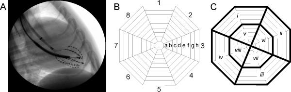
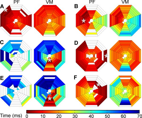
Methods: A multielectrode basket catheter was deployed in the left ventricle of eight beagles. External defibrillation shocks were delivered with a ramp-up protocol after SDVF (20 seconds) and LDVF (150 seconds). Earliest VM and PF activations were identified after the highest energy shock that failed to terminate VF and the successful shock.
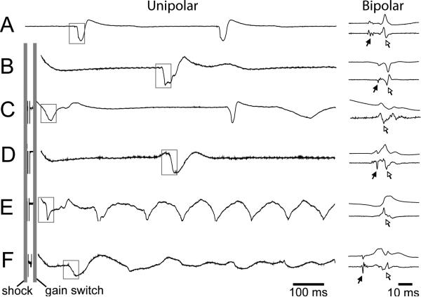
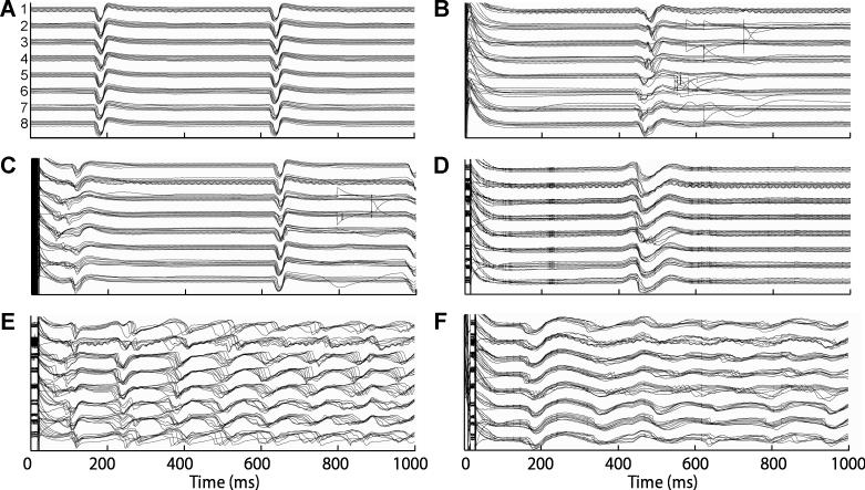
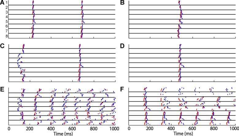
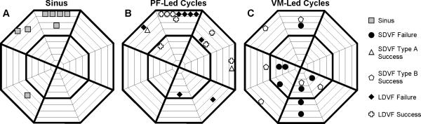
Results: Defibrillation was successful after 36 +/- 12 and 181 +/- 14 seconds for SDVF and LDVF, respectively. The time after shock delivery until earliest activation was detected for failed shocks and was significantly longer after LDVF (138.7 +/- 24.1 ms) than after SDVF (75.6 +/- 8.7 ms). Earliest postshock activation after SDVF typically initiated in the VM (14 of 16 episodes), while it always initiated in the PF (16 of 16 episodes) after LDVF. Sites of earliest activity during sinus rhythm correlated with sites of earliest postshock activation for PF-led cycles but not for VM-led cycles.
Conclusion: Earliest recorded postshock activation is in the Purkinje system after LDVF but not after SDVF. This difference raises the possibility that the optimal defibrillation strategy is different for SDVF and LDVF.
Copyright 2010 Heart Rhythm Society. Published by Elsevier Inc. All rights reserved.
Figures

Similar articles
-
Transmural recording of shock potential gradient fields, early postshock activations, and refibrillation episodes associated with external defibrillation of long-duration ventricular fibrillation in swine.Heart Rhythm. 2008 Nov;5(11):1599-606. doi: 10.1016/j.hrthm.2008.08.019. Epub 2008 Aug 28. Heart Rhythm. 2008. PMID: 18984539 Free PMC article.
-
Effect of flunarizine on defibrillation outcomes and early refibrillation in a canine model of prolonged ventricular fibrillation.Exp Physiol. 2019 Nov;104(11):1630-1637. doi: 10.1113/EP087068. Epub 2019 Oct 9. Exp Physiol. 2019. PMID: 31465138 Free PMC article.
-
The effects of acute amiodarone on short- and long-duration ventricular defibrillation threshold in canines.J Cardiovasc Pharmacol. 2011 Oct;58(4):432-8. doi: 10.1097/FJC.0b013e318228a50c. J Cardiovasc Pharmacol. 2011. PMID: 21709582
-
[Ventricular fibrillation and electric defibrillation].Dtsch Med Wochenschr. 1989 Jan 27;114(4):145-9. doi: 10.1055/s-2008-1066568. Dtsch Med Wochenschr. 1989. PMID: 2644115 Review. German. No abstract available.
-
Mechanisms of VF maintenance: wandering wavelets, mother rotors, or foci.Heart Rhythm. 2009 Mar;6(3):405-15. doi: 10.1016/j.hrthm.2008.11.005. Epub 2008 Nov 8. Heart Rhythm. 2009. PMID: 19251220 Free PMC article. Review.
Cited by
-
Rotor stability separates sustained ventricular fibrillation from self-terminating episodes in humans.J Am Coll Cardiol. 2014 Jun 24;63(24):2712-21. doi: 10.1016/j.jacc.2014.03.037. Epub 2014 Apr 30. J Am Coll Cardiol. 2014. PMID: 24794115 Free PMC article.
-
Three-dimensional mechanisms of increased vulnerability to electric shocks in myocardial infarction: altered virtual electrode polarizations and conduction delay in the peri-infarct zone.J Physiol. 2012 Sep 15;590(18):4537-51. doi: 10.1113/jphysiol.2012.229088. Epub 2012 May 14. J Physiol. 2012. PMID: 22586222 Free PMC article.
-
Undersensing of ventricular fibrillation by a biventricular implantable cardioverter-defibrillator: What is the cause and the troubleshooting?J Arrhythm. 2019 Feb 22;35(2):276-278. doi: 10.1002/joa3.12170. eCollection 2019 Apr. J Arrhythm. 2019. PMID: 31007793 Free PMC article. No abstract available.
-
Mechanisms of defibrillation.Annu Rev Biomed Eng. 2010 Aug 15;12:233-58. doi: 10.1146/annurev-bioeng-070909-105305. Annu Rev Biomed Eng. 2010. PMID: 20450352 Free PMC article. Review.
-
Spatiotemporal Progression of Early Human Ventricular Fibrillation.JACC Clin Electrophysiol. 2017 Dec 11;3(12):1437-1446. doi: 10.1016/j.jacep.2017.04.009. Epub 2017 Aug 2. JACC Clin Electrophysiol. 2017. PMID: 29238755 Free PMC article.
References
-
- Valenzuela TD, Roe DJ, Nichol G, et al. Outcomes of Rapid Defibrillation by Security Officers after Cardiac Arrest in Casinos. N Engl J Med. 2000;343:1206–1209. - PubMed
-
- Dosdall DJ, Fast V, Ideker RE. Mechanisms of defibrillation. In: Zipes DP, Jalife J, editors. Cardiac electrophysiology : from cell to bedside. 5th ed Saunders; Philadelphia: 2009. pp. 499–508.
-
- Chattipakorn N, Fotuhi PC, Ideker RE. Prediction of defibrillation outcome by epicardial activation patterns following shocks near the defibrillation threshold. J Cardiovasc Electrophysiol. 2000;11:1014–21. - PubMed
Publication types
MeSH terms
Grants and funding
LinkOut - more resources
Full Text Sources
Other Literature Sources
