

The E2 ubiquitin-conjugating enzymes direct polyubiquitination to preferred lysines
- PMID: 20061386
- PMCID: PMC2838281
- DOI: 10.1074/jbc.M109.089003
The E2 ubiquitin-conjugating enzymes direct polyubiquitination to preferred lysines
Abstract
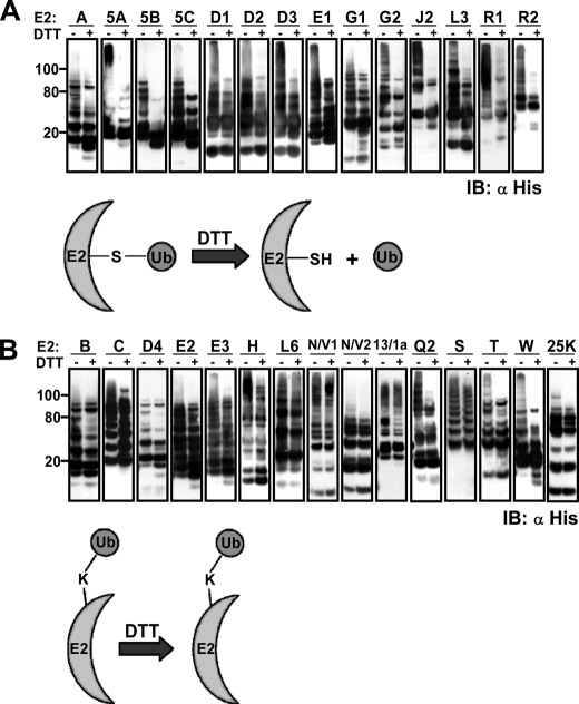
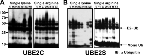
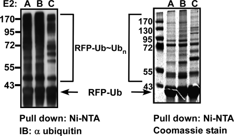
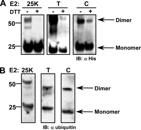
The ubiquitin-proteasome pathway plays a crucial role in many cellular processes by degrading substrates tagged by polyubiquitin chains, linked mostly through lysine 48 of ubiquitin. Although polymerization of ubiquitin via its six other lysine residues exists in vivo as part of various physiological pathways, the molecular mechanisms that determine the type of polyubiquitin chains remained largely unknown. We undertook a systematic, in vitro, approach to evaluate the role of E2 enzymes in determining the topology of polyubiquitin. Because this study was performed in the absence of an E3 enzyme, our data indicate that the E2 enzymes are capable of directing the ubiquitination process to distinct subsets of ubiquitin lysines, depending on the specific E2 utilized. Moreover, our findings are in complete agreement with prior analyses of lysine preference assigned to certain E2s in the context of E3 (in vitro and in vivo). Finally, our findings support the rising notion that the functional unit of E2 is a dimer. To our knowledge, this is the first systematic indication for the involvement of E2 enzymes in specifying polyubiquitin chain assembly.
Figures

Similar articles
-
Two different classes of E2 ubiquitin-conjugating enzymes are required for the mono-ubiquitination of proteins and elongation by polyubiquitin chains with a specific topology.Biochem J. 2008 Feb 1;409(3):723-9. doi: 10.1042/BJ20071338. Biochem J. 2008. PMID: 18042044
-
Certain pairs of ubiquitin-conjugating enzymes (E2s) and ubiquitin-protein ligases (E3s) synthesize nondegradable forked ubiquitin chains containing all possible isopeptide linkages.J Biol Chem. 2007 Jun 15;282(24):17375-86. doi: 10.1074/jbc.M609659200. Epub 2007 Apr 10. J Biol Chem. 2007. PMID: 17426036
-
Direct catalysis of lysine 48-linked polyubiquitin chains by the ubiquitin-activating enzyme.J Biol Chem. 2007 Dec 28;282(52):37454-60. doi: 10.1074/jbc.M705242200. Epub 2007 Oct 19. J Biol Chem. 2007. PMID: 17951259
-
The family of ubiquitin-conjugating enzymes (E2s): deciding between life and death of proteins.FASEB J. 2010 Apr;24(4):981-93. doi: 10.1096/fj.09-136259. Epub 2009 Nov 25. FASEB J. 2010. PMID: 19940261 Review.
-
Structural basis of generic versus specific E2-RING E3 interactions in protein ubiquitination.Protein Sci. 2019 Oct;28(10):1758-1770. doi: 10.1002/pro.3690. Epub 2019 Aug 23. Protein Sci. 2019. PMID: 31340062 Free PMC article. Review.
Cited by
-
Arabidopsis membrane-anchored ubiquitin-fold (MUB) proteins localize a specific subset of ubiquitin-conjugating (E2) enzymes to the plasma membrane.J Biol Chem. 2011 Apr 29;286(17):14913-21. doi: 10.1074/jbc.M110.158808. Epub 2011 Feb 23. J Biol Chem. 2011. PMID: 21345795 Free PMC article.
-
The anti-inflammatory drug BAY 11-7082 suppresses the MyD88-dependent signalling network by targeting the ubiquitin system.Biochem J. 2013 May 1;451(3):427-37. doi: 10.1042/BJ20121651. Biochem J. 2013. PMID: 23441730 Free PMC article.
-
UBE2B is implicated in myofibrillar protein loss in catabolic C2C12 myotubes.J Cachexia Sarcopenia Muscle. 2016 Jun;7(3):377-87. doi: 10.1002/jcsm.12060. Epub 2015 Nov 19. J Cachexia Sarcopenia Muscle. 2016. PMID: 27239408 Free PMC article.
-
Key genes and regulatory networks involved in the initiation, progression and invasion of colorectal cancer.Future Sci OA. 2018 Jan 24;4(3):FSO278. doi: 10.4155/fsoa-2017-0108. eCollection 2018 Mar. Future Sci OA. 2018. PMID: 29568567 Free PMC article.
-
Dietary effects on the retina of hamsters.FASEB J. 2025 Mar 31;39(6):e70451. doi: 10.1096/fj.202403390R. FASEB J. 2025. PMID: 40099968 Free PMC article.
References
-
- Goldberg A. L. (1995) Science 268, 522–523 - PubMed
-
- Hershko A., Ciechanover A. (1998) Annu. Rev. Biochem. 67, 425–479 - PubMed
-
- Hochstrasser M. (1996) Annu. Rev. Genet 30, 405–439 - PubMed
-
- Köhler A., Cascio P., Leggett D. S., Woo K. M., Goldberg A. L., Finley D. (2001) Mol. Cell 7, 1143–1152 - PubMed
Publication types
MeSH terms
Substances
LinkOut - more resources
Full Text Sources
Other Literature Sources
Molecular Biology Databases
