

Physiological numbers of CD4+ T cells generate weak recall responses following influenza virus challenge
- PMID: 20061406
- PMCID: PMC2826830
- DOI: 10.4049/jimmunol.0901427
Physiological numbers of CD4+ T cells generate weak recall responses following influenza virus challenge
Abstract
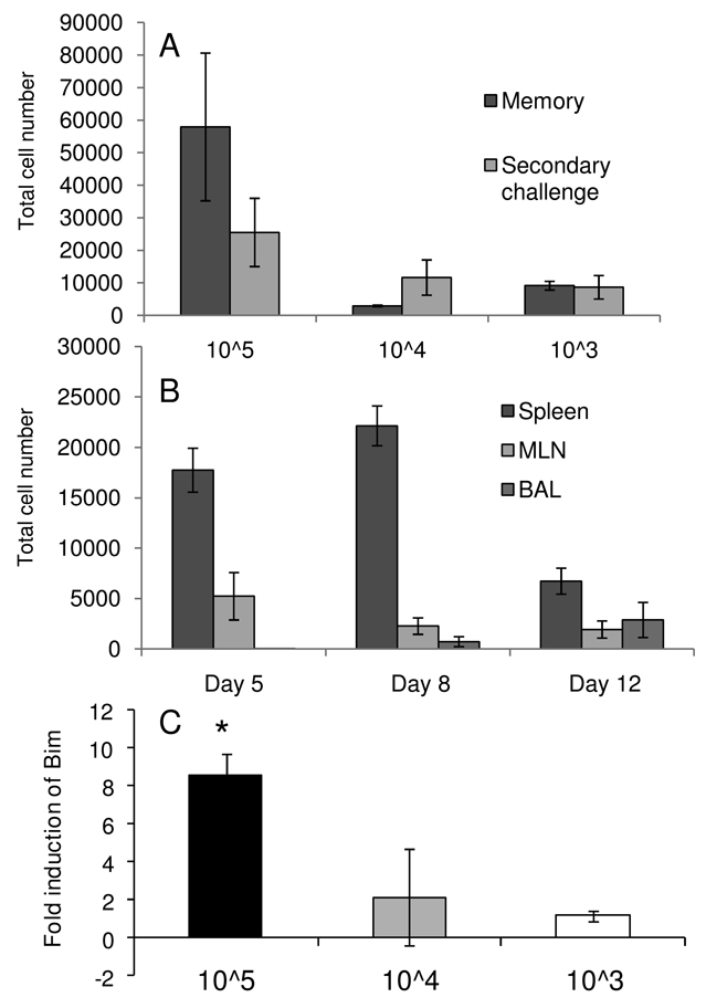
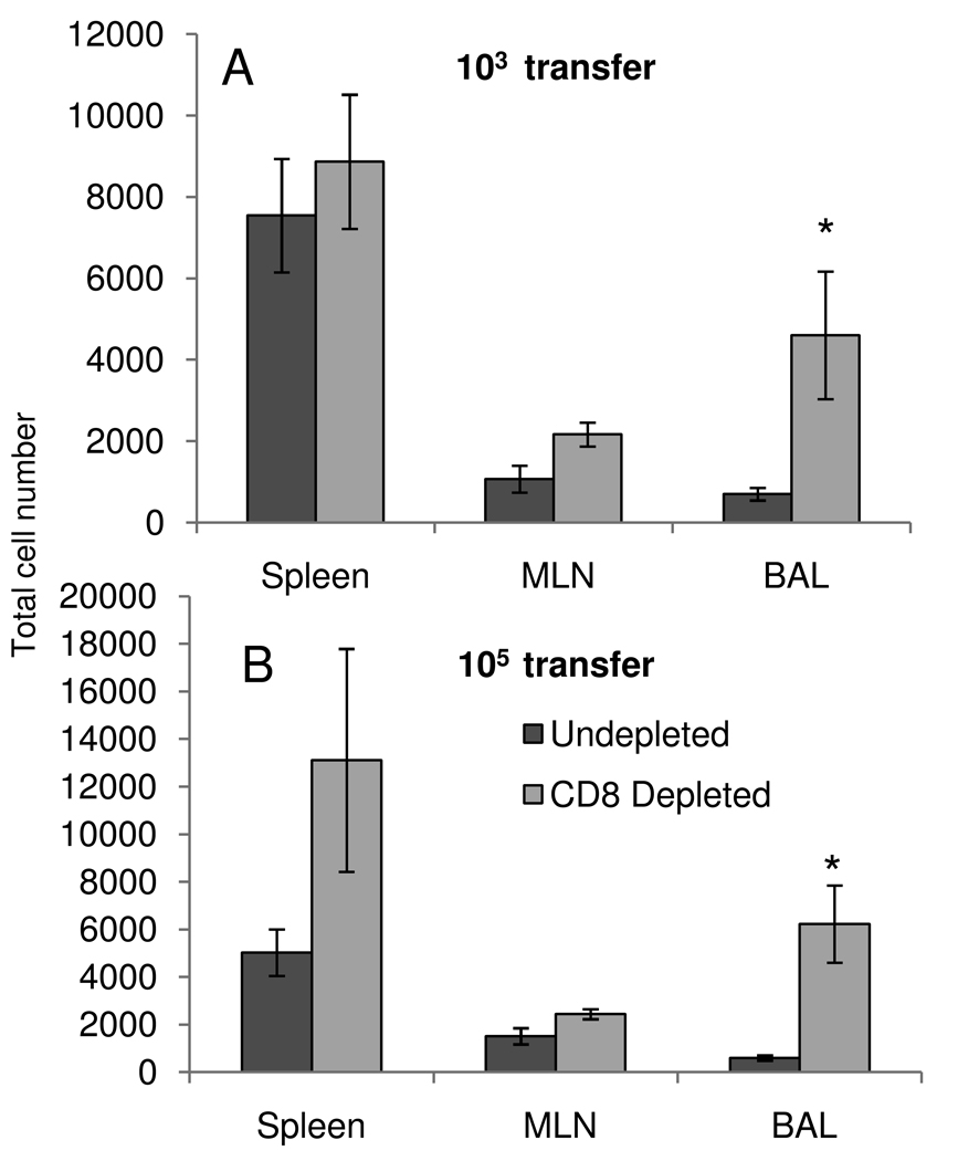
Naive and recall CD4(+) T cell responses were probed with recombinant influenza A viruses incorporating the OVA OT-II peptide. The extent of OT-II-specific CD4(+) T cell expansion was greater following primary exposure, with secondary challenge achieving no significant increase in numbers, despite higher precursor frequencies. Adoptive transfer experiments with OT-II TCR-transgenic T cells established that the predominant memory set is CD62L(hi), whereas the CD62L(lo) precursors make little contribution to the recall response. Unlike the situation described by other investigators, in which the transfer of very large numbers of in vitro-activated CD4 effectors can modify the disease process, providing CD62L(hi) or CD62L(lo) OT-II-specific T cells at physiological levels neither enhanced virus clearance nor altered clinical progression. Some confounding effects of the transgenic model were observed, with decreasing primary expansion efficiency correlating with greater numbers of transferred cells. This was associated with increased levels of mRNA for the proapoptotic molecule Bim in cells recovered following high-dose transfer. However, even with very low numbers of transferred cells, memory T cells did not expand significantly following secondary challenge. A similar result was recorded in mice primed and boosted to respond to an endogenous IA(b)-restricted epitope derived from the influenza virus hemagglutinin glycoprotein. Depletion of CD8(+) T cells during secondary challenge generated an increased accumulation of OT-II-specific T cells but only at the site of infection. Taken together, significant expansion was not a feature of these secondary influenza-specific CD4 T cell responses and the recall of memory did not enhance recovery.
Figures


Similar articles
-
Memory precursor phenotype of CD8+ T cells reflects early antigenic experience rather than memory numbers in a model of localized acute influenza infection.Eur J Immunol. 2011 Mar;41(3):682-93. doi: 10.1002/eji.201040625. Epub 2011 Jan 25. Eur J Immunol. 2011. PMID: 21264852
-
Homogenization of TCR repertoires within secondary CD62Lhigh and CD62Llow virus-specific CD8+ T cell populations.J Immunol. 2008 Jun 15;180(12):7938-47. doi: 10.4049/jimmunol.180.12.7938. J Immunol. 2008. PMID: 18523257
-
Memory CD4 T cells direct protective responses to influenza virus in the lungs through helper-independent mechanisms.J Virol. 2010 Sep;84(18):9217-26. doi: 10.1128/JVI.01069-10. Epub 2010 Jun 30. J Virol. 2010. PMID: 20592069 Free PMC article.
-
Recalling the Future: Immunological Memory Toward Unpredictable Influenza Viruses.Front Immunol. 2019 Jul 2;10:1400. doi: 10.3389/fimmu.2019.01400. eCollection 2019. Front Immunol. 2019. PMID: 31312199 Free PMC article. Review.
-
CD4+ T-cell memory: generation and multi-faceted roles for CD4+ T cells in protective immunity to influenza.Immunol Rev. 2006 Jun;211:8-22. doi: 10.1111/j.0105-2896.2006.00388.x. Immunol Rev. 2006. PMID: 16824113 Free PMC article. Review.
Cited by
-
Heterosubtypic T-Cell Immunity to Influenza in Humans: Challenges for Universal T-Cell Influenza Vaccines.Front Immunol. 2016 May 19;7:195. doi: 10.3389/fimmu.2016.00195. eCollection 2016. Front Immunol. 2016. PMID: 27242800 Free PMC article. Review.
-
Competition within the virus-specific CD4 T-cell pool limits the T follicular helper response after influenza infection.Immunol Cell Biol. 2016 Sep;94(8):729-40. doi: 10.1038/icb.2016.42. Epub 2016 Apr 22. Immunol Cell Biol. 2016. PMID: 27101922
-
Tolerance induction in memory CD4 T cells is partial and reversible.Immunology. 2021 Jan;162(1):68-83. doi: 10.1111/imm.13263. Epub 2020 Oct 27. Immunology. 2021. PMID: 32931017 Free PMC article.
-
The establishment of surrogates and correlates of protection: Useful tools for the licensure of effective influenza vaccines?Hum Vaccin Immunother. 2018 Mar 4;14(3):647-656. doi: 10.1080/21645515.2017.1413518. Epub 2018 Jan 16. Hum Vaccin Immunother. 2018. PMID: 29252098 Free PMC article. Review.
-
Influenza infection fortifies local lymph nodes to promote lung-resident heterosubtypic immunity.J Exp Med. 2021 Jan 4;218(1):e20200218. doi: 10.1084/jem.20200218. J Exp Med. 2021. PMID: 33005934 Free PMC article.
References
-
- Christensen JP, Bartholdy C, Wodarz D, Thomsen AR. Depletion of CD4+ T cells precipitates immunopathology in immunodeficient mice infected with a noncytocidal virus. J Immunol. 2001;166:3384–3391. - PubMed
-
- Su HC, Cousens LP, Fast LD, Slifka MK, Bungiro RD, Ahmed R, Biron CA. CD4+ and CD8+ T cell interactions in IFN-gamma and IL-4 responses to viral infections: requirements for IL-2. J Immunol. 1998;160:5007–5017. - PubMed
Publication types
MeSH terms
Substances
Grants and funding
LinkOut - more resources
Full Text Sources
Research Materials
