. 2010 Feb 15;184(4):1675-9.
doi: 10.4049/jimmunol.0903539. Epub 2010 Jan 8.

Cutting edge: spontaneous development of IL-17-producing gamma delta T cells in the thymus occurs via a TGF-beta 1-dependent mechanism

Affiliations

Cutting edge: spontaneous development of IL-17-producing gamma delta T cells in the thymus occurs via a TGF-beta 1-dependent mechanism

Jeong-su Do et al. J Immunol. .

Abstract

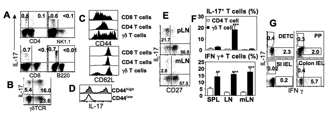
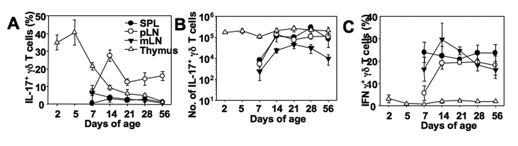
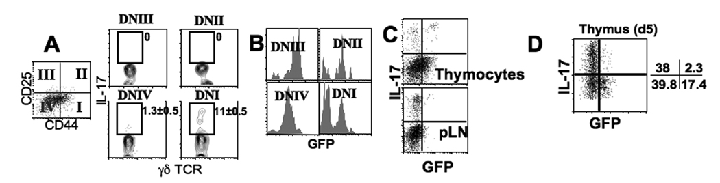
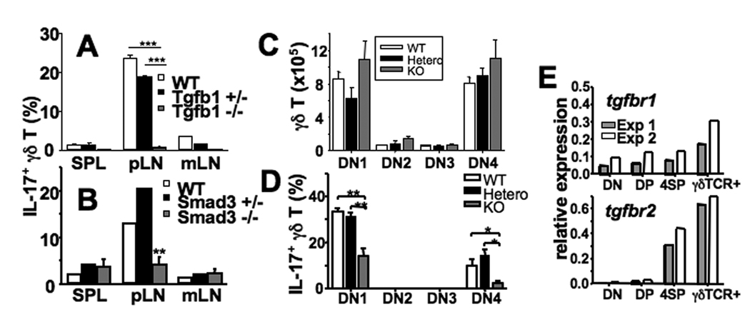
In naive animals, gammadelta T cells are innate sources of IL-17, a potent proinflammatory cytokine mediating bacterial clearance as well as autoimmunity. However, mechanisms underlying the generation of these cells in vivo remain unclear. In this study, we show that TGF-beta1 plays a key role in the generation of IL-17(+) gammadelta T cells and that it mainly occurs in the thymus particularly during the postnatal period. Interestingly, IL-17(+) gammadelta TCR(+) thymocytes were mainly CD44(high)CD25(low) cells, which seem to derive from double-negative 4 gammadelta TCR(+) cells that acquired CD44 and IL-17 expression. Our findings identify a novel developmental pathway during which IL-17-competent gammadelta T cells arise in the thymus by a TGF-beta1-dependent mechanism.


Figures

Figure 1
Figure 1. pLN γδ T cells preferentially express IL-17 following activation
(A) LN cells were ex vivo stimulated with PMA/ionomycin and subsequently stained for CD4, NK1.1, CD8, B220, and IL-17. (B) LN cells stimulated as above were stained for B220, CD4, CD8, IL-17, and γδ TCR. Shown is γδ T cell IL-17 expression by non-B/non-T cells. (C) γ δ, CD4, and CD8 T cells from the indicated tissues were examined for the surface expression of CD44 and CD62L. (D) IL-17 expression of CD44low and CD44high γδ T cells (spleen and pLN) was examined by intracellular staining. (E) γδ T cells from the indicated tissues were stimulated and stained for IL-17 and CD27. (F) Proportions of IFNγ- and of IL-17-producing γδ T cells (and CD4 T cells) in the indicated tissues were examined. Shown is the mean ± SD (n=4). (G) Cells isolated from the indicated tissues were subsequently stimulated with PMA/Ionomycin, and cytokine production was determined by flow cytometric analysis. Shown are cytokine profiles of γδ TCR+ gated cells. All the experiments were repeated more than twice and similar results were observed. **, p<0.01, ***, p<0.001.
Figure 2
Figure 2. IL-17 expression of γδ T cells in mice of different ages
(A–C) Spleen, pLN, mLN, and thymic cells from mice at the indicated ages were stimulated as described in Figure 1 were stained for IL-17, IFNγ, and γδ TCR. Proportions of IL-17- (A) and of IFNγ- (C) expressing γδ T cells were examined. (B) Total numbers of IL-17-expressing γδ T cells were determined by flow analysis. Shown are the mean ± SD (n = 3–4).
Figure 3
Figure 3. IL-17-expressing thymic γδ T cells
(A) Adult thymic cells stimulated as described in Figure 1 were stained for CD44, CD25, γδ TCR, and IL-17. Quadrant plot represents γδ TCR+ gated thymocytes. IL-17 expression of each subset (based on CD44 and CD25 expression) was indicated. (B) Thymic cells from adult Rag2p-GFP Tg mice were stained for γδ TCR, CD44, and CD25. GFP expression of each DN subset is shown. The results are representative of 4 individually tested mice. (C) Thymocytes and pLN cells from adult Rag2p-GFP Tg mice were stimulated and IL-17/GFP expression of γδ T cells was examined. (D) Thymic cells from 5-day old Rag2p-GFP mice were stimulated and stained for γδ TCR, CD44, CD25, and IL-17. Shown is GFP expression profiles of CD44highCD25low γδ T cells. The results are representative of 4 individually tested mice.
Figure 4
Figure 4. TGFβ1 controls γδ T cell IL-17 expression
(A and B) γδ T cell cytokine production from Tgfβ1−/− (A), Smad3−/− (B), and littermate control mice was determined. Shown are the mean ± SD of 4~6 individually tested mice. (C and D) Thymic cells from 11-day old Tgfβ1−/− and littermate control mice were examined for DN subset distribution based on CD44 and CD25 expression (C), or stimulated and IL-17 expression of γδ TCR+ thymocytes in each DN subset was examined (D). Shown is the mean ± SD of 4 individually tested mice. (E) FACS sorted DN γδ TCR- (DN), DP, CD4 SP (4SP), and DN γδ TCR+ thymocytes were examined for Tgfβr1 and Tgfβr2 expression by real time PCR. *, p<0.05; **, p<0.01; ***, p<0.001.

References

    1. Carding SR, Egan PJ. Gammadelta T cells: functional plasticity and heterogeneity. Nat Rev Immunol. 2002;2:336–345. - PubMed
    1. Shibata K, Yamada H, Hara H, Kishihara K, Yoshikai Y. Resident Vdelta1+ gammadelta T cells control early infiltration of neutrophils after Escherichia coli infection via IL-17 production. J Immunol. 2007;178:4466–4472. - PubMed
    1. Roark CL, Simonian PL, Fontenot AP, Born WK, O'Brien RL. gammadelta T cells: an important source of IL-17. Curr Opin Immunol. 2008;20:353–357. - PMC - PubMed
    1. Stark MA, Huo Y, Burcin TL, Morris MA, Olson TS, Ley K. Phagocytosis of apoptotic neutrophils regulates granulopoiesis via IL-23 and IL-17. Immunity. 2005;22:285–294. - PubMed
    1. Jensen KD, Su X, Shin S, Li L, Youssef S, Yamasaki S, Steinman L, Saito T, Locksley RM, Davis MM, Baumgarth N, Chien YH. Thymic selection determines gammadelta T cell effector fate: antigen-naive cells make interleukin-17 and antigen-experienced cells make interferon gamma. Immunity. 2008;29:90–100. - PMC - PubMed

Publication types

MeSH terms

Substances