Strategic distribution of protective proteins within bran layers of wheat protects the nutrient-rich endosperm
- PMID: 20061449
- PMCID: PMC2832279
- DOI: 10.1104/pp.109.149864
Strategic distribution of protective proteins within bran layers of wheat protects the nutrient-rich endosperm
Abstract
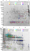
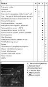
Bran from bread wheat (Triticum aestivum 'Babbler') grain is composed of many outer layers of dead maternal tissues that overlie living aleurone cells. The dead cell layers function as a barrier resistant to degradation, whereas the aleurone layer is involved in mobilizing organic substrates in the endosperm during germination. We microdissected three defined bran fractions, outer layers (epidermis and hypodermis), intermediate fraction (cross cells, tube cells, testa, and nucellar tissue), and inner layer (aleurone cells), and used proteomics to identify their individual protein complements. All proteins of the outer layers were enzymes, whose function is to provide direct protection against pathogens or improve tissue strength. The more complex proteome of the intermediate layers suggests a greater diversity of function, including the inhibition of enzymes secreted by pathogens. The inner layer contains proteins involved in metabolism, as would be expected from live aleurone cells, but this layer also includes defense enzymes and inhibitors as well as 7S globulin (specific to this layer). Using immunofluorescence microscopy, oxalate oxidase was localized predominantly to the outer layers, xylanase inhibitor protein I to the xylan-rich nucellar layer of the intermediate fraction and pathogenesis-related protein 4 mainly to the aleurone. Activities of the water-extractable enzymes oxalate oxidase, peroxidase, and polyphenol oxidase were highest in the outer layers, whereas chitinase activity was found only in assays of whole grains. We conclude that the differential protein complements of each bran layer in wheat provide distinct lines of defense in protecting the embryo and nutrient-rich endosperm.
Figures


Similar articles
-
Proteome evolution of wheat (Triticum aestivum L.) aleurone layer at fifteen stages of grain development.J Proteomics. 2015 Jun 18;123:29-41. doi: 10.1016/j.jprot.2015.03.008. Epub 2015 Apr 2. J Proteomics. 2015. PMID: 25841591
-
Cell Wall Proteome of Wheat Grain Endosperm and Outer Layers at Two Key Stages of Early Development.Int J Mol Sci. 2019 Dec 29;21(1):239. doi: 10.3390/ijms21010239. Int J Mol Sci. 2019. PMID: 31905787 Free PMC article.
-
Distinct metabolic changes between wheat embryo and endosperm during grain development revealed by 2D-DIGE-based integrative proteome analysis.Proteomics. 2016 May;16(10):1515-36. doi: 10.1002/pmic.201500371. Epub 2016 Apr 27. Proteomics. 2016. PMID: 26968330
-
Proteomes of the barley aleurone layer: A model system for plant signalling and protein secretion.Proteomics. 2011 May;11(9):1595-605. doi: 10.1002/pmic.201000656. Epub 2011 Mar 23. Proteomics. 2011. PMID: 21433287 Review.
-
Wheat bran layers: composition, structure, fractionation, and potential uses in foods.Crit Rev Food Sci Nutr. 2024;64(19):6636-6659. doi: 10.1080/10408398.2023.2171962. Epub 2023 Feb 2. Crit Rev Food Sci Nutr. 2024. PMID: 36728922 Review.
Cited by
-
Polyphenol oxidase as a biochemical seed defense mechanism.Front Plant Sci. 2014 Dec 10;5:689. doi: 10.3389/fpls.2014.00689. eCollection 2014. Front Plant Sci. 2014. PMID: 25540647 Free PMC article.
-
Delineation of Inheritance Pattern of Aleurone Layer Colour Through Chemical Tests in Rice.Rice (N Y). 2017 Nov 21;10(1):48. doi: 10.1186/s12284-017-0187-9. Rice (N Y). 2017. PMID: 29164348 Free PMC article.
-
Mixed-Linkage Glucan Is the Main Carbohydrate Source and Starch Is an Alternative Source during Brachypodium Grain Germination.Int J Mol Sci. 2023 Apr 6;24(7):6821. doi: 10.3390/ijms24076821. Int J Mol Sci. 2023. PMID: 37047802 Free PMC article.
-
The Dead Can Nurture: Novel Insights into the Function of Dead Organs Enclosing Embryos.Int J Mol Sci. 2018 Aug 19;19(8):2455. doi: 10.3390/ijms19082455. Int J Mol Sci. 2018. PMID: 30126259 Free PMC article. Review.
-
The cereal starch endosperm development and its relationship with other endosperm tissues and embryo.Protoplasma. 2015 Jan;252(1):33-40. doi: 10.1007/s00709-014-0687-z. Epub 2014 Aug 16. Protoplasma. 2015. PMID: 25123370 Review.
References
-
- Antoine C, Peyron S, Lullien-Pellerin V, Abecassis J, Rouau X. (2004) Wheat bran tissue fractionation using biochemical markers. J Cereal Sci 39: 387–393
-
- Antoine C, Peyron S, Mabille F, Lapierre C, Bouchet B, Abecassis J, Rouau X. (2003) Individual contribution of grain outer layers and their cell wall structure to the mechanical properties of wheat bran. J Agric Food Chem 51: 2026–2033 - PubMed
-
- Blein JP, Coutos-Thévenot P, Marion D, Ponchet M. (2002) From elicitins to lipid-transfer proteins: a new insight in cell signalling involved in plant defence mechanisms. Trends Plant Sci 7: 293–296 - PubMed
-
- Bonnin E, Daviet S, Gebruers K, Delcour JA, Goldson A, Juge N, Saulnier L. (2005) Variation in the levels of the different xylanase inhibitors in grain and flour of 20 French wheat cultivars. J Cereal Sci 41: 375–379
-
- Bradford MM. (1976) A rapid and sensitive method for the quantitation of microgram quantities of protein utilizing the principle of protein-dye binding. Anal Biochem 72: 248–254 - PubMed
Publication types
MeSH terms
Substances
LinkOut - more resources
Full Text Sources

