. 2010 Mar;192(5):1193-200.
doi: 10.1128/JB.01391-09. Epub 2010 Jan 8.

The chemoreceptor dimer is the unit of conformational coupling and transmembrane signaling

Affiliations

The chemoreceptor dimer is the unit of conformational coupling and transmembrane signaling

Divya N Amin et al. J Bacteriol. 2010 Mar.

Abstract

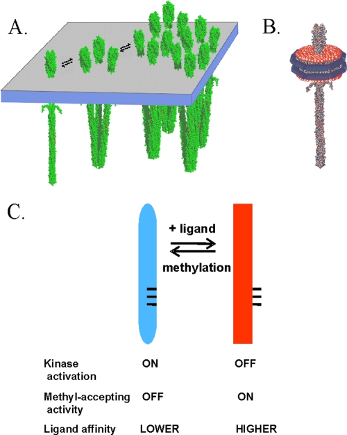
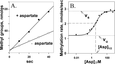
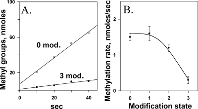
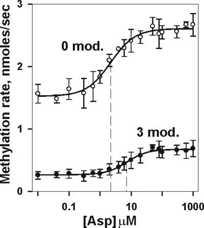
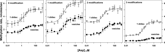
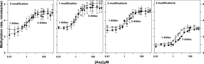
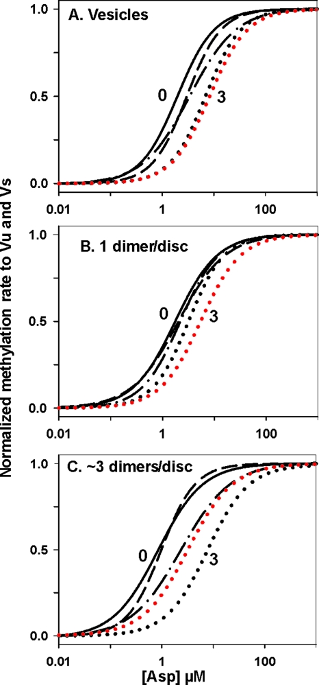
Transmembrane chemoreceptors are central components in bacterial chemotaxis. Receptors couple ligand binding and adaptational modification to receptor conformation in processes that create transmembrane signaling. Homodimers, the fundamental receptor structural units, associate in trimers and localize in patches of thousands. To what degree do conformational coupling and transmembrane signaling require higher-order interactions among dimers? To what degree are they altered by such interactions? To what degree are they inherent features of homodimers? We addressed these questions using nanodiscs to create membrane environments in which receptor dimers had few or no potential interaction partners. Receptors with many, few, or no interaction partners were tested for conformational changes and transmembrane signaling in response to ligand occupancy and adaptational modification. Conformation was assayed by measuring initial rates of receptor methylation, a parameter independent of receptor-receptor interactions. Coupling of ligand occupancy and adaptational modification to receptor conformation and thus to transmembrane signaling occurred with essentially the same sensitivity and magnitude in isolated dimers as for dimers with many neighbors. Thus, we conclude that the chemoreceptor dimer is the fundamental unit of conformational coupling and transmembrane signaling. This implies that in signaling complexes, coupling and transmembrane signaling occur through individual dimers and that changes between dimers in a receptor trimer or among trimer-based signaling complexes are subsequent steps in signaling.


Figures

FIG. 1.
FIG. 1.
Chemoreceptors. (A) Cartoon of interactions of membrane-embedded chemoreceptors showing a homodimer, a trimer of dimers, and a patch of chemoreceptors. (B) Cartoon of a nanodisc with a single receptor dimer inserted in the plug of the lipid bilayer. (C) Diagram of the chemoreceptor conformational equilibrium.
FIG. 2.
FIG. 2.
Initial rates of Tar methylation enhanced by aspartate. (A) Time courses of methylation in the absence and presence of a saturating concentration (1 mM) of the Tar ligand aspartate. (B) Initial rate of methylation as a function of aspartate concentration. Data like the data shown in panel A were collected for Tar(QEQE) embedded in native membrane vesicles at the indicated concentrations of aspartate. The curve is a fit of the data to a simple dose-response relationship (see Materials and Methods). Dotted lines labeled vu and vs indicate the initial rates of methylation in the absence of aspartate and in the presence of a saturating concentration of the ligand, respectively. Dashed lines indicate the aspartate concentration, [Asp]1/2, at which enhancement of the initial rate of methylation is half-maximal.
FIG. 3.
FIG. 3.
Initial rates of Tar methylation reduced by adaptational modification. (A) Time courses of methylation for Tar with 0 or 3 (QEQQ) methyl-accepting sites modified by the introduction of glutamine, a functional analog of a methyl ester. (B) Initial rate of methylation as a function of adaptational modification. Data like the data shown in panel A were collected for Tar embedded in native membrane vesicles carrying 0, 1 (QEEE), 2 (QEQE), or 3 (QEQQ) glutamines at the 4 methyl-accepting sites of that chemoreceptor. The curve is drawn to guide the eye.
FIG. 4.
FIG. 4.
Initial rates of methylation as a function of adaptational modification and ligand concentration. Rates were determined for Tar embedded in native membrane vesicles carrying 0 or 3 (QEQQ) glutamines at the indicated aspartate concentrations. The graph shows the means ± standard deviations (error bars) for ≥3 independent experiments. The curves are fits of the data to a simple dose-response relationship (see Materials and Methods). The vertical dashed line shows the respective values for [Asp]1/2.
FIG. 5.
FIG. 5.
Effects of ligand and adaptational modification on initial rate of methylation persist in chemoreceptor dimers isolated from interacting neighbors. Experiments, data, and curves are as described in the legend to Fig. 4, using the Tar with patterns of modification described in the legend to Fig. 3. The figures compare Tar inserted in native membrane vesicles (vesicles) in which each receptor dimer had many potentially interacting neighbors and inserted in nanodiscs at 1 dimer/disc (1 d/disc) a condition with no potentially interacting neighbors.
FIG. 6.
FIG. 6.
Effects of ligand and adaptational modification on initial rate of methylation for chemoreceptors with no or few interacting neighbors. Dose-response curves of initial rates of methylation as a function of aspartate concentration for chemoreceptor Tar with no (1 dimer/disc) or few (∼3 dimers/disc) potentially interacting neighboring receptors were determined as described in the legend to Fig. 4.
FIG. 7.
FIG. 7.
Effects of chemoreceptor adaptational modification on functional affinity for ligand as influenced by the presence of potentially interacting neighboring receptors. Tar with many potentially interacting neighbors (A), no potentially interacting neighbors (B), or few potentially interacting neighbors (C) were compared for effects of adaptational modification on the dose-response relationship of the Tar ligand aspartate and initial rate of methylation. To facilitate comparison of the dose-response relationships for receptors with 0 (solid black line), 1 (QEEE; dashed line), 2 (QEQE; dot-dash line), and 3 (QEQQ [black dotted line] and QQEQ [red dotted line]) modifications, the curves shown in Fig. 5 and 6 were normalized to vu and to vs and displayed without the data points from which they had been derived.

Similar articles

Cited by

References

    1. Barnakov, A., L. Barnakova, and G. Hazelbauer. 1998. Comparison in vitro of a high- and a low-abundance chemoreceptor of Escherichia coli: similar kinase activation but different methyl-accepting activities. J. Bacteriol. 180:6713-6718. - PMC - PubMed
    1. Bayburt, T. H., and S. G. Sligar. 2003. Self-assembly of single integral membrane proteins into soluble nanoscale phospholipid bilayers. Protein Sci. 12:2476-2481. - PMC - PubMed
    1. Besschetnova, T. Y., D. J. Montefusco, A. E. Asinas, A. L. Shrout, F. M. Antommattei, and R. M. Weis. 2008. Receptor density balances signal stimulation and attenuation in membrane-assembled complexes of bacterial chemotaxis signaling proteins. Proc. Natl. Acad. Sci. U. S. A. 105:12289-12294. - PMC - PubMed
    1. Boldog, T., S. Grimme, M. Li, S. G. Sligar, and G. L. Hazelbauer. 2006. Nanodiscs separate chemoreceptor oligomeric states and reveal their signaling properties. Proc. Natl. Acad. Sci. U. S. A. 103:11509-11514. - PMC - PubMed
    1. Boldog, T., M. Li, and G. L. Hazelbauer. 2007. Using Nanodiscs to create water-soluble transmembrane chemoreceptors inserted in lipid bilayers. Methods Enzymol. 423:317-335. - PubMed

Publication types

LinkOut - more resources