. 2010 Mar;192(6):1498-510.
doi: 10.1128/JB.01167-09. Epub 2010 Jan 8.

PepD participates in the mycobacterial stress response mediated through MprAB and SigE

Affiliations

PepD participates in the mycobacterial stress response mediated through MprAB and SigE

Mark J White et al. J Bacteriol. 2010 Mar.

Abstract

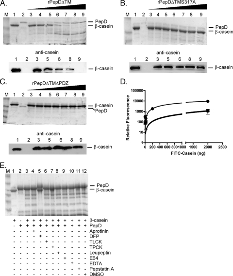
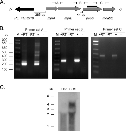
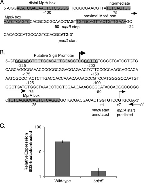
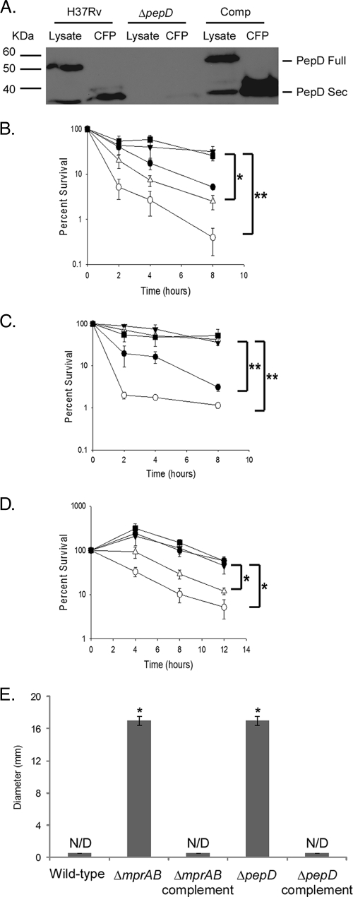
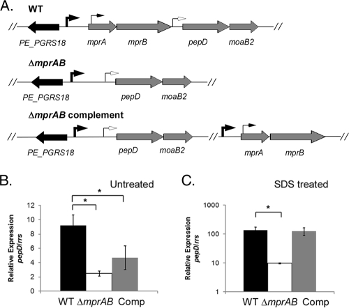
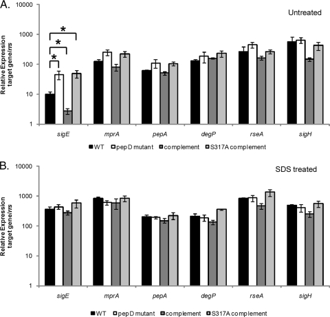
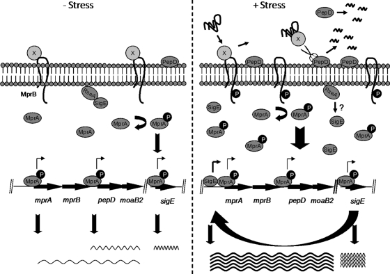
Currently, one-third of the world's population is believed to be latently infected with Mycobacterium tuberculosis. The mechanisms by which M. tuberculosis establishes latent infection remain largely undefined. mprAB encodes a two-component signal transduction system required by M. tuberculosis for aspects of persistent infection. MprAB regulates a large and diverse group of genetic determinants in response to membrane stress, including the extracytoplasmic function (ECF) sigma factor sigE and the HtrA-like serine protease pepD. Recent studies have demonstrated that PepD functions as both a protease and chaperone in vitro. In addition, inactivation of pepD alters the virulence of M. tuberculosis in a mouse model system of infection. Here, we demonstrate that PepD plays an important role in the stress response network of Mycobacterium mediated through MprAB and SigE. In particular, we demonstrate that the protease activity of PepD requires the PDZ domain, in addition to the catalytic serine at position 317. pepD expression initiates from at least three promoters in M. tuberculosis, including one that is regulated by SigE and is located upstream of the mprA coding sequence. Deletion of pepD or mprAB in Mycobacterium smegmatis and M. tuberculosis alters the stress response phenotypes of these strains, including increasing sensitivity to SDS and cell wall antibiotics and upregulating the expression of stress-responsive determinants, including sigE. Taking these data together, we hypothesize that PepD utilizes its PDZ domain to recognize and process misfolded proteins at the cell membrane, leading to activation of the MprAB and SigE signaling pathways and subsequent establishment of a positive feedback loop that facilitates bacterial adaptation.


Figures

FIG. 1.
FIG. 1.
rPepDΔTM exhibits proteolytic activity against the artificial substrate β-casein. Protease assays were carried out with the artificial substrate β-casein and rPepDΔTM (A), rPepDΔTMS317A (B), or rPepDΔTMΔPDZ (C) for 16 h at 37°C. The reaction mixtures contained 1 μg β-casein alone (lanes 1), 500 ng rPepD variant alone (lanes 2), or 1 μg β-casein incubated with increasing concentrations (67 to 500 ng) of rPepD variants (lanes 3 to 9). The reaction products were resolved by SDS-PAGE and visualized by either Coomassie staining or Western blotting using a polyclonal antibody against casein. (D) The cleavage of β-casein by rPepD variants was quantified using 41.5 μg FITC-labeled casein as a substrate and measuring the relative fluorescence of reaction products (loss of FITC quenching) as a function of the rPepD concentration. The rPepDΔTMS317A and rPepDΔTMΔPDZ variants exhibited approximately 10% of the activity of rPepDΔTM. •, rPepDΔTM; ▪, rPepDΔTMS317A; ⧫, rPepDΔTMΔPDZ. The error bars indicate standard errors. (E) The specificity of rPepDΔTM-mediated β-casein cleavage was determined by the inclusion of various protease inhibitors in the reaction mixture. The reaction mixtures contained 1 μg β-casein alone (lane 1), 500 ng rPepDΔTM alone (lane 2), or 1 μg β-casein and 500 ng rPepDΔTM (lane 3) incubated with 1.5 mM aprotinin (lane 4), 5 mM DFP (lane 5), 27 mM TLCK (lane 6), 28 mM TPCK (lane 7), 23 mM leupeptin (lane 8), 1 mM E64 (lane 9), 50 mM EDTA (lane 10), 1 mM pepstatin A (lane 11), or DMSO (lane 12). Inhibition was observed in the reaction mixture containing DFP. Lanes M, molecular weight marker.
FIG. 2.
FIG. 2.
Transcriptional organization of the pepD locus in M. tuberculosis. (A) Chromosomal organization of pepD and surrounding genes in M. tuberculosis H37Rv. The primers used to determine the operon structure and the probe generated for Northern blot hybridization are shown. (B) Agarose gels depicting RT-PCRs with primer sets spanning intergenic regions. Lanes: M, marker lane; +RT, RNA treated with reverse transcriptase; −RT, RNA treated without reverse transcriptase; +, genomic DNA; −, no template. (C) Northern blot of RNA samples obtained from either untreated (Unt) or SDS-treated M. tuberculosis H37Rv cultures.
FIG. 3.
FIG. 3.
Characterization of the pepD promoter region. (A and B) Genomic depiction of pepD (A) and mprA (B) upstream regions, including MprA binding sites (gray boxes), determined transcriptional start sites (arrows), and annotated or predicted translational start and stop sites (boldface letters). Note that one of the transcriptional start sites for mprA is downstream of the translational start site previously annotated. The location of the primer set used to measure expression from the distal mprA promoter is also shown by arrows. (C) The wild type and the ΔsigE M. tuberculosis mutant were grown in the absence of SDS or were exposed to 0.05% SDS for 90 min, and the relative amounts of expression from the distal mprA promoter were determined. Expression levels were normalized to those of the 16S ribosomal gene, rrs, and are expressed as relative expression from cultures exposed to SDS versus untreated cultures. The results represent the means ± standard errors of the mean (SEM) from three independent experiments performed in triplicate.
FIG. 4.
FIG. 4.
Characterization of mycobacterial ΔmprAB and ΔpepD mutants. (A) Western blot analysis of cell lysates and culture filtrate protein (CFP) from wild-type, ΔpepD, and complemented M. tuberculosis strains probed with the α-PepD antibody. (B and C) Sensitivities of wild-type, mutant, and complemented M. smegmatis derivatives (B) or M. tuberculosis derivatives (C) to SDS as determined by broth sensitivity assays. The results are expressed as percent survival between untreated cultures and cultures treated with 0.1% or 0.4% SDS, respectively. (D) Survival of M. smegmatis derivatives after exposure to 200 μg/ml d-cycloserine expressed as percent survival between untreated and treated cultures. Survival for broth sensitivity assays was determined by plating aliquots onto 7H10 agar medium and assaying them for CFU. (E) Sensitivities of M. smegmatis derivatives to cefuroxime as determined by disc diffusion assay. The results are expressed as zones of inhibition (N/D, not detected). ▪, wild-type; ○, ΔmprAB; ▵, ΔpepD; •, ΔmprAB complement; ▴, ΔpepD complement. The results represent the means ± SEM from three independent experiments performed in triplicate. *, P < 0.05; **, P < 0.01.
FIG. 5.
FIG. 5.
Polarity resulting from the ΔmprAB mutation. (A) Genomic organization of the mprA-mprB-pepD-moaB2 locus in M. tuberculosis H37Rv (wild type [WT], the ΔmprAB mutant, and complemented strains. The arrows depict the identified transcriptional start sites in the region. (B and C) Wild-type M. tuberculosis, the ΔmprAB mutant, and the ΔmprAB complemented strain were grown without SDS (B) or were exposed to 0.05% SDS for 90 min (C). The relative amounts of pepD in the strains were compared. The expression levels were normalized to that of the 16S ribosomal gene, rrs. The results represent the means ± SEM from three independent experiments performed in triplicate. *, P < 0.05.
FIG. 6.
FIG. 6.
Impact of pepD deletion on M. tuberculosis gene expression in the absence or presence of SDS stress. (A and B) Wild-type M. tuberculosis, the ΔpepD mutant, the ΔpepD complemented strain, and the ΔpepD S317A complemented strain were grown in the absence of stress (A) or were exposed to 0.05% SDS for 90 min (B). The relative amounts of sigE, mprA, degP, pepA, rseA, or sigH expression in the strains were then compared. The expression level for each gene under each condition was normalized to that of the 16S ribosomal gene, rrs. The results represent the means ± SEM from three independent experiments performed in triplicate. *, P < 0.05.
FIG. 7.
FIG. 7.
Model of the activation of the MprAB system. Under nonstress conditions (left), an unidentified protein repressor (protein X) binds to MprB, keeping it in an inactive conformation and resulting in basal-level expression of mprAB transcription. In the presence of membrane stress (right), misfolded proteins accumulate in the extracytoplasmic space, resulting in the release of protein X from MprB and activation of the MprAB signaling pathway, including upregulation of sigE and subsequent induction of mprAB-pepD-moaB. PepD is secreted into the extracytoplasmic space between the cell membrane and the cell wall, where it functions to degrade and/or refold damaged substrate targets. Following proteolysis, the PDZ domain of PepD is removed in an autocatalytic event, resulting in release of PepD into the culture supernatant. Wavy lines indicate transcripts, with darker lines indicating higher expression lavels.

References

    1. Alba, B. M., H. J. Zhong, J. C. Pelayo, and C. A. Gross. 2001. degS (hhoB) is an essential Escherichia coli gene whose indispensable function is to provide sigma (E) activity. Mol. Microbiol. 40:1323-1333. - PubMed
    1. Anonymous. 2008. Cycloserine. Tuberculosis (Edinb.) 88:100-101. - PubMed
    1. Anonymous. 2008. Global tuberculosis control. Surveillance, planning, financing. World Health Organization, Geneva, Switzerland.
    1. Baranova, N., and H. Nikaido. 2002. The baeSR two-component regulatory system activates transcription of the yegMNOB (mdtABCD) transporter gene cluster in Escherichia coli and increases its resistance to novobiocin and deoxycholate. J. Bacteriol. 184:4168-4176. - PMC - PubMed
    1. Bardarov, S., S. Bardarov, Jr., M. S. Pavelka, Jr., V. Sambandamurthy, M. Larsen, J. Tufariello, J. Chan, G. Hatfull, and W. R. Jacobs, Jr. 2002. Specialized transduction: an efficient method for generating marked and unmarked targeted gene disruptions in Mycobacterium tuberculosis, M. bovis BCG and M. smegmatis. Microbiology 148:3007-3017. - PubMed

Publication types

MeSH terms

Substances